Biomedical Engineering Reference
In-Depth Information
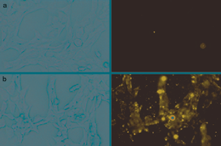
Fig. 5
In vitro
interaction of rhodamine-PE labeled PEG-PE micelles with 4 T1 cells. Left panel
shows the bright field and right panel shows the fluorescent microscopy of 4 T1 cells treated with
rhodamine-PE labeled micelles. (
a
), rhodamine-PE :PEG
750
-PE micelles; (
b
), rhodamine-PE:
PEG
750
-PE: TATp-PEG
1000
-PE micelles. Magnification ×40 objective (Modified from Sawant and
Torchilin
2009
)
apoptosis was observed using the TUNEL assay with a DNA fragmentation kit.
Very few TUNEL-positive cells were observed in tumors injected with free pacli-
taxel and paclitaxel-loaded micelles (Fig.
6c
). However, significant apoptotic cell
death was observed in tumors treated with paclitaxel-loaded TATp-bearing
micelles (Fig.
6d
).
Another application of CPPs involves the labeling of cells with semiconductor
nanocrystals or quantum dots (QDs). QDs are now more popular than standard
fluorophores for the study of tumor pathophysiology since they are photostable,
more robust stable light emitters, and relatively insensitive to the wavelength of
the excitation. They are also capable of distinguishing tumor vessels from both the
perivascular cells and the matrix, with concurrent imaging. QDs trapped within
PEG-PE micelles bearing a TATp-PEG-PE linker were used to label mouse
endothelial cells
in vitro
. For
in vivo
tracking, bone marrow-derived progenitor
cells labeled with TATp-bearing QD-containing micelles
ex vivo
, were injected in
mice bearing a tumor in a cranial window model. It was possible to track the
movement of labeled progenitor cells to the tumor endothelium, that may provide
a path towards the understanding of the fine details of tumor neovascularization
(Stroh et al.
2005
).
Search WWH ::

Custom Search