Biomedical Engineering Reference
In-Depth Information
2
Intracellular Delivery of Lipid-Core Micelles
with Cationic Lipids
Polymeric micelles cannot diffuse through the cell membrane but rather are internalized
by endocytosis. Detailed reviews of the endocytotic pathways and endocytosis of
nanocarriers can be found in (Conner and Schmid
2003
; Mukherjee et al.
1997
;
Bareford and Swaan
2007
). Following cell uptake, micelles are contained within
acidic endosomes and are further directed to various transport pathways including
fusion with lysosomes or exocytosis. Therefore, it is necessary to further improve
the efficiency of drug-loaded micelles by enhancement of their intracellular deliv-
ery to compensate for excessive drug degradation in lysosomes as a result of the
endocytosis-mediated capture of micelles by cells.
PEG-PE micelles carry a net negative charge (Lukyanov and Torchilin
2004
),
which can hinder their internalization by cells. Modification of PEG-PE micelles
with positively charged lipids may improve the uptake of drug-loaded micelles by
cells. Such positively charged micelles could also more readily escape from endo-
somes and enter the cytoplasm. To test these ideas, we have prepared paclitaxel-
loaded micelles from mixture of PEG-PE and positively charged Lipofectin
®
lipids
(LL) (Wang et al.
2005
). The intracellular fate of paclitaxel-loaded PEG-PE/LL
micelles and micelles prepared without the addition of the LL was investigated by
fluorescence microscopy with BT-20 breast adenocarcinoma cells. Both fluorescently-
labeled PEG-PE and PEG-PE/LL micelles were endocytosed by BT-20 cells
(Fig.
1
). However, with PEG-PE/LL micelles, endosomes appeared to be partially
disrupted and released drug-loaded micelles into the cell cytoplasm, a result of the
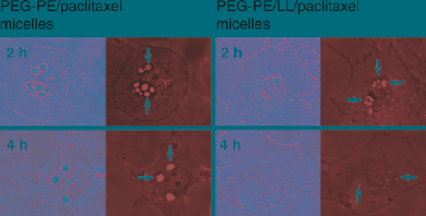
Fig. 1
Microscopy of BT-20 cells incubated with PEG-PE/ paclitaxel micelles and PEG-PE/
LL/paclitaxel micelles for 2 and 4 h. Bright-field (
left images
in each pair) and fluorescence
(
right images
in each pair).
Arrows
indicate fluorescent endosomes in cells incubated with
PEG-PE/paclitaxel micelles for 2 h; partially degraded endosomes in cells incubated with
PEG-PE/LL/paclitaxel micelles for 2 h; punctuate fluorescent structures in cells incubated
with PEG-PE/LL/paclitaxel micelles for 4 h; larger (fused) endosomes in cells incubated with
PEG-PE/paclitaxel micelles for 4 h (Modified from Torchilin
2005a
)
Search WWH ::

Custom Search