Biology Reference
In-Depth Information
[18,43]
in the rat still remains the best and most widely characterized model for
reproducing widespread injury to the axonal structures in the white matter. Early
markers of axonal damage, the amyloid precursor protein (APP) and heavy chain
neurofilament (200 kDa), almost identically overlap in immunohistochemical
staining, where the axons present with swelling and retraction bulbs. Preliminary
experiments in our laboratory have demonstrated abundant axonal damage follow-
ing diffuse traumatic axonal injury, particularly within the corpus callosum and the
pyramidal tract of the brainstem, which are subject to high forces during the trau-
matic impact. This axonal damage is exacerbated when a 30-minute hypoxic insult
is superimposed on the TBI (
Figure 10.2
). Povlishock's group has investigated this
model over several years and found that axonal dysfunction is a process that begins
early after trauma and slowly progresses, leading to impaired retrograde transport,
ionic dysregulation, axonal swelling, and ultimately axonal disconnection
[44]
. The
underlying mechanisms include axonal Ca
2
influx and increased permeability of the
axolemma, followed by mitochondrial dysfunction and activation of apoptotic cas-
pases. Although axonal perturbation can be reversed, when the point of no return is
reached, it proceeds into total axotomy. Compared to focal TBI, diffuse axonal injury
does not involve a massive accumulation of blood leukocytes, but only infiltration
of macrophages/microglia in discrete regions such as subarachnoid spaces and the
(A)
(B)
(C)
(D)
(E)
(F)
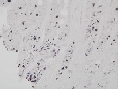
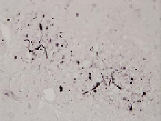
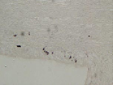
Figure 10.2
Representative microscope images demonstrating axonal damage in the rat
brain at 1 day following diffuse traumatic axonal injury (TAI). Axonal damage was detected
using immunohistochemistry for neurofilament (200 kDa heavy chain), which is vulnerable
to compaction and accumulation in axons following TAI. Distinct patterns of damage, in
the form of retraction bulbs and axonal swelling, are present in the pyramidal tracts of
the brainstem (A, B, C) and corpus callosum (D, E, F) in rats that have undergone TAI in
combination with hypoxia (A, D) or TAI alone (B, E). Note that the concomitant insult of
hypoxia results in a far greater level of axonal injury than that observed in TAI alone. This
severity of injury is evident when taken in the context of sham-treated animals (C, F), which
are completely devoid of neurofilament accumulation. Scale bar 100 m.