Agriculture Reference
In-Depth Information
be inserted one or more claws; the ambulacrum
on leg I may be small or absent. In some groups
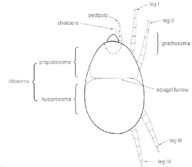
of mites, the idiosoma is subdivided by a sejugal
furrow into an anterior propodosoma and a pos-
terior hysterosoma
(Fig. 166),
each bearing two
pairs of legs. Mites within the superfamily
Eriophyoidea (order Prostigmata) possess just
two pairs of legs, both of which arise from the
propodosoma. Ocelli, when present, are located
on the propodosoma.
The body of a mite often bears more or less
sclerotized plates (shields) and these are useful
for distinguishing between various groups.
Identification of mites, however, usually requires
high-powered
detailed examination of setae on the body and
appendages (chaetotaxy) is of particular impor-
tance but is a specialist task and beyond the
scope of the present work. Features of the shield
which overlies the propodosoma of eriophyid
mites (in the present work termed the prodorsal
shield - also widely known as the cephalo-
thoracic shield, the dorsal shield and the
propodosomal shield), and the number and
orientation of the setae arising from it, are of
considerable taxonomic significance; although
brief mention of the more gross features is made
in the specific descriptions (see Part II, p. 255
et
seq.),
their full appreciation requires the use of a
scanning electron microscope.
microscopical
examination;
INTERNAL FEATURES
nervous tissue (the central nerve mass) around
the pharynx. The alimentary canal includes a
pharynx, an elongated oesophagus, a mid-gut
(ventriculus) and a hindgut, the anterior part
of which may include several Malpighian-like
tubules.
Apart from bearing the pedipalps and the
mouthparts, the gnathosoma is little more than a
tube through which the foregut passes into the
idiosoma. It does not contain a brain, the nervous
system of a mite being located within the
propodosoma and, typically, forming a ring of
Fig. 166
General structure of a mite.