Chemistry Reference
In-Depth Information
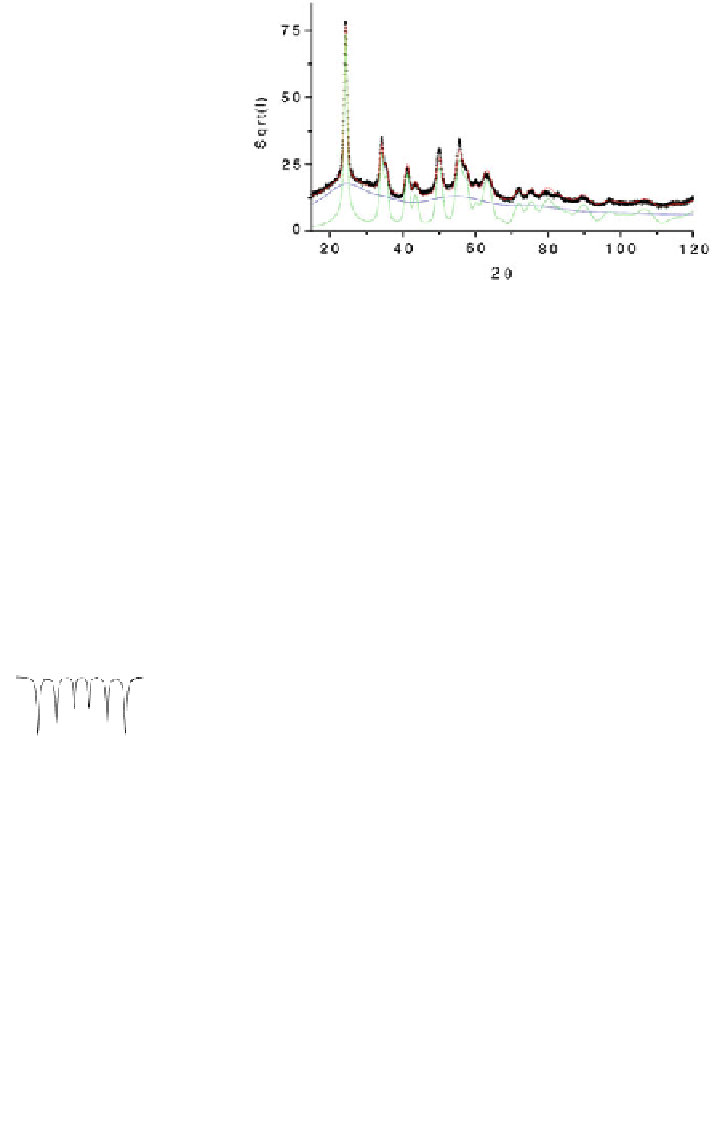
Fig. 4.22 Typical X-ray
pattern of milled FeF
3
and its
decomposition into two
components attributed to
crystalline grains and grain
boundaries [
165
]
part of Bragg peaks. The X-ray diffraction pattern modelling gave successfully rise to the
presence of two main components with broadened peaks, the proportions of which are
rather equivalent. The first component with well resolved peaks is attributed to rhom-
bohedral nanocrystalline and spherical grains with a size of about 15 nm diameter while
the second component corresponding to the low part is characteristics of structural
coherent diffraction domain of about 0.4 nm (equivalent to the size of an FeF
6
octahedral
unit): it is thus assigned to grain boundaries resulting from a packing of octahedral units.
Series of Mössbauer spectra were recorded for different grinding times and
grinding energy at different temperatures and in presence of external magnetic
field. The modelling does take into account simultaneously the corresponding
hyperfine structures [
166
]. Examples of spectra and corresponding hyperfine field
distributions are given in Fig.
4.23
recorded at 300 and 4.2 K on r-FeF
3
in the
100
100
80
60
50
40
20
60
0
0
30
40
50
0
10
20
30
40
50
2,0
50
25
1,0
25
60
0
0
0
10
20
30
40
0 0 0 0
30
40
50
50
50
2
25
25
1
0
0
60
0
0
10
20
30
40
0 0 0 0
30
40
50
50
50
2
25
1
25
0
0
0
0
10
20
30
40
0 0 0 0
30
40
50
60
50
2
50
25
1
25
0
0
60
0
0
10
20
30
40
0 0 0 0
30
40
50
-10
-5
0
5
10
-12
-6
0
6
12
mm/s
mm/s
Fig. 4.23
Mössbauer
spectra
recorded
at
300 K
(left)
and
4.2 K
(right)
on
as-prepared
crystalline FeF
3
(top) and ground for 0.25, 1, 8 and 16 h (top to bottom)[
166
]