Chemistry Reference
In-Depth Information
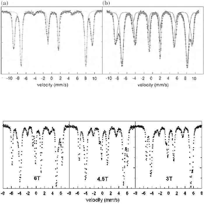
Fig. 3.49 External-field (6 T) spectra at RT of well-crystallized (a) and poorly crystallized
(b) maghemite. Note the presence of lines 2 and 5 as an indication of spin canting
Fig. 3.50 Mössbauer spectra of the greigite-smythite sample at 80 K in different external
magnetic fields showing the separation of the greigite lines
A mineral-related example was the use of external field measurements in the
study of greigite [
148
]. The RT spectrum of a greigite-smythite sediment sample
has been reproduced in
Sect. 3.4.1
(Fig.
3.19
). This spectrum has been fitted with
two sextets from greigite, three sextets from smythite and a siderite doublet. As
shown, the spectrum fit seems at first sight to be straightforward, but that was not
at all the case. At that moment, the hyperfine fields on tetrahedral and octahedral
sites of greigite were not well known and the purpose of that study was to
determine the temperature behavior of the hyperfine fields. The only way to tackle
the problem consisted of measuring the sample at 80 K in applied fields of various
strengths. Because greigite is ferrimagnetic the external field separates its A- en
B-site spectrum (Fig.
3.50
), whereas the lines of smythite, being ferromagnetic
shift more to center of the spectrum. Extrapolation for the different fields yielded
the values of the hyperfine fields at 80 K and the respective isomer shifts.