Information Technology Reference
In-Depth Information
Table 1 DE parameters used
for leukocytes detection in
medical images
m
F
CR
NI
20
0.25
0.80
200
Step 1:
Segment the WBC
'
s using the DEM algorithm (described in
4.1
)
Step 2:
Get the edge map from the segmented image
Step 3:
Start the ellipse detector based in DE over the edge map while saving best ellipses
(Sect.
3
)
Step 4:
Define parameter values for each ellipse that identify the WBC
'
s
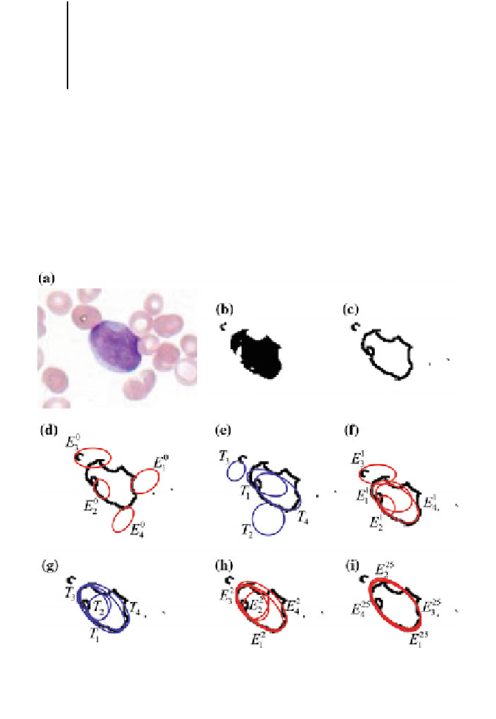
4.3 Numerical Example
'
In order to present the algorithm
s step-by-step operation, a numerical example has
been set by applying the proposed method to detect a single leukocyte lying inside
of a simple image. Figure
7
a shows the image used in the example. After applying
Fig. 7 Detection numerical example: a the image used as example. b Segmented image. c Edge
map. d Initial particles E
0
. e Trial elements T produced by the DE operators. f New population E
1
.
g Trial elements produced considering E
1
as input population. h New population E
2
. i Final
particle configuration after 25 iterations


Search WWH ::

Custom Search