Environmental Engineering Reference
In-Depth Information
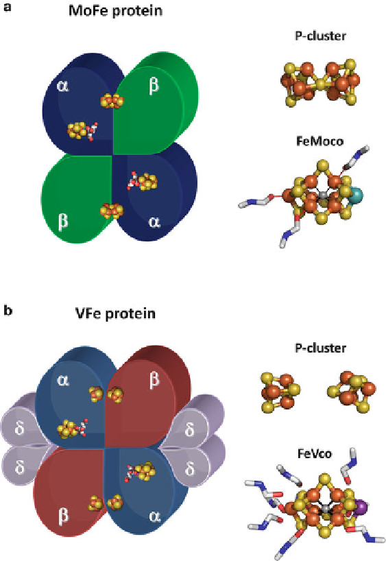
Figure 8 Schematic representations of the MoFe (a,
left
) and VFe (b,
left
) proteins,
EXAFS-derived models of the P-clusters in the MoFe (a,
top right
) and VFe (b,
top right
) proteins,
and EXAFS-derived models of the NMF-extracted FeMoco (a,
bottom right
) and FeVco (b,
bottom right
). The MoFe protein is an
ʱ
2
ʲ
2
heterotetramer (a,
left
); whereas the VFe protein is
an
4
heterooctamer (b,
left
). The P-cluster of the MoFe protein has an [Fe
8
S
7
] topology
(a,
top right
); whereas the P-cluster of the VFe protein consists of paired [Fe
4
S
4
] clusters (b,
top
right
). The isolated FeVco (b,
bottom right
) resembles the isolated FeMoco (a,
bottom right
)in
core structure, although it is more extended in structure and bound with more NMF molecules.
Clusters are shown in ball-and-stick presentations; NMF molecules are shown as sticks. Atoms are
colored as follows: Fe, orange; S, yellow; V, dark purple; Mo, cyan; C, white; N, blue; O, red.
PYMOL was used to generate this figure, with coordinates of the P-cluster and the FeMoco in
the crystal structure of the MoFe protein (PDB entry 1M1N) modified on the basis of the Fe-edge
EXAFS fits.
ʱ
2
ʲ
ʴ
2
Search WWH ::

Custom Search