Environmental Engineering Reference
In-Depth Information
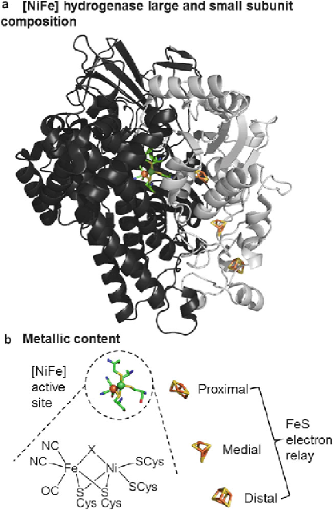
Figure 6 The structure of a [NiFe] hydrogenase. (a) Depicts the minimal functional unit for activity,
consisting of the large (dark grey ribbon) protein subunit encasing the bimetallic active site and the
small (light grey ribbon) protein subunit encasing the FeS relay. Image was generated using the
Escherichia coli
hydrogenase-1 structure, PDB code 3UQY. (b) Amore detailed view of the metallic
centers. Color code: yellow depicts sulfur; blue depicts nitrogen; bright red depicts oxygen; green
ball depicts Ni, green sticks depict hydrocarbon; orange-red ball and sticks depict iron.
Search WWH ::

Custom Search