Graphics Reference
In-Depth Information
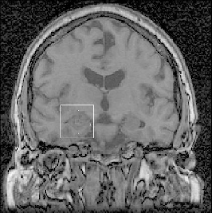
Fig. 10.22.
The usage of multiple sources and sinks to control the evolution of the
GMS algorithm for the fully automated segmentation of the hippocampus in the
human brain (from [64]).
(a)
(b)
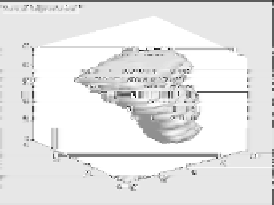
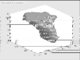
Fig. 10.23.
Comparison of manual and automatic segmentation of the hippocampus
in the human brain. Image (a) is a manual segmentation by a clinician that required
about 2 hours of labeling and (b) is a fully-automated segmentation via GMS using
multiple sources and sinks positioned by cross-validated training on labeled images,
which required just 2 minutes of computation (from [64]).
Search WWH ::

Custom Search