Biology Reference
In-Depth Information
a
Initial
Control w/o KO
2
10
µ
M KO
2
20
µ
M KO
2
30
µ
M KO
2
TMRM
CM-DCF
b
0.09
0.06
0.03
0.00
0
10
20
30
KO
2
(
M)
µ
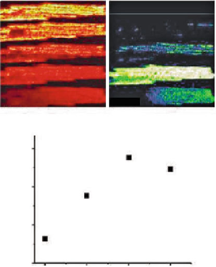
Fig. 5.9 Mitochondrial O
2
.
and
ΔΨ
m
in response to increased exogenous O
2
.
Myocytes were
loaded with TMRM (100 nM) and CM-H
2
DCFDA (2
μ
M) for at least 20 min and imaged using
two-photon laser scanning fluorescence microscopy. After loading, the excess dye was washed out
and the cells were briefly superfused with a permeabilizing solution (saponin) (Aon et al. 2007).
After permeabilization, the myocytes were continuously perfused with an intracellular solution
containing GSH:GSSG at a ratio of 300:1. The TMRM was included in the medium to avoid
depletion of the probe during depolarization-repolarization cycles. (a) The TMRM and CM-DCF
images of a permeabilized cardiomyocyte at time zero after loading and before (
top row image
)or
after permeabilization and 5 min imaging under control conditions (Control,
second row
) or the
presence of KO
2
,anO
2
.
donor (10
μ
M, third row; 20
μ
M, fourth row; 30
μ
M, fifth row), after
3 min equilibration in each case. RIRR-mediated
m
depolarization without a permeability
transition occurs at the two lower concentrations, while loss of the CM-DCF probe (~500 MW)
from the mitochondrial matrix due to PTP opening occurs at 30
ΔΨ
MKO
2
.(b) The rates of O
2
.
accumulation as a function of KO
2
concentration. Slopes were calculated when the linear rate of
change of the CM-DCF signal stabilized under each condition. Reproduced from Zhou, Aon,
Almas, Cortassa, Winslow, O'Rourke (2010) PLoS Computational Biology 6(1): e1000657.
doi:10.1371/journal.pcbi.1000657
μ
concentration, from 10 to 20
ΔΨ
m
depolarization and
increased the rate of mitochondrial O
2
.
accumulation (Fig.
5.9b
). Exposure of
the cell to 30
μ
M, elicited progressive
μ
MKO
2
induced an irreversible collapse of
ΔΨ
m
, accompanied by the
Search WWH ::

Custom Search