Biomedical Engineering Reference
In-Depth Information
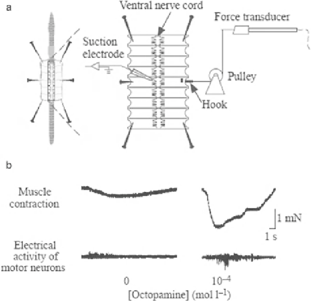
Fig. 6.4
Circular muscle contraction induced by octopamine. (
a
) Diagram of a semi-intact
preparation. A small thread which is connected to a force transducer via a pulley is hooked in the
middle part of the dissected body wall. The electrical activity of motor neurons was recorded
simultaneously from the cut end of the fi rst segmental nerve in the same segment using a glass
suction electrode. (
b)
The simultaneous recording of muscle contraction and electrical activity
from projecting nerves. In control preparations, electrical activity was only seen at the onset of a
contraction. At 10
-4
M octopamine, electrical activity occurred throughout much of the contraction
period (modifi ed from Mizutani et al.
2002
)
2004
). The simultaneous measurement of muscle contraction and neuronal activity
from the segmental nerve cord is shown in Fig.
6.4a
. Previously, we confi rmed that the
neuronal activity from the left and right segmental nerve cord is coincident during fi c-
tive locomotion, and octopamine induces the huge contraction of the circular muscles
with more frequent neuronal activity (Fig.
6.4b
). However, the motor neurons and
interneurons underlying fi ctive locomotion were still unknown at that time; therefore,
we tried to identify such neurons using optical imaging techniques.
The development of various imaging techniques and fl uorescent dyes has enabled
researchers to visualize the mobilization of various substances in neurons. We have
various choices for Ca indicators, for example, Fura-2, Fluo-3 and Fluo-4, and
Calcium Green-1, and also several kinds of fl uorescent protein-based Ca sensors,
including Cameleon (Miyawaki et al.
1997
). We previously examined Ca wave