Biomedical Engineering Reference
In-Depth Information
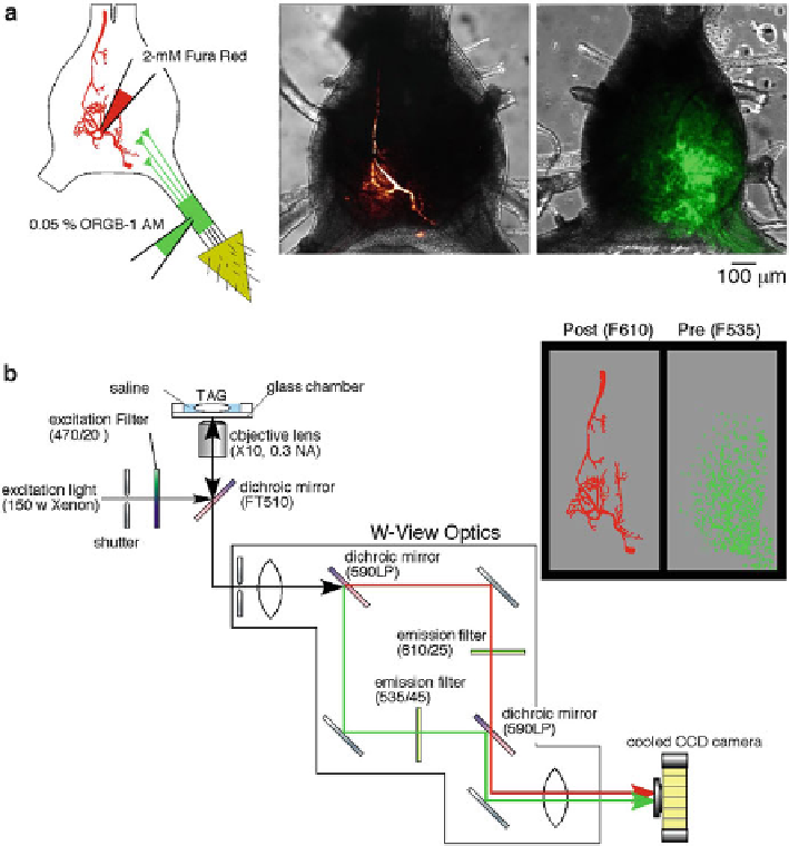
Fig. 5.4
Experimental setup for simultaneous imaging of Ca
2+
signals in the pre- and postsynaptic
neurons in the cricket cercal sensory system. (
a
) Diagrams showing the methods for selective load-
ing of the different Ca
2+
indicators. A solution containing OGB-1 AM was pressure injected
through a glass micropipette into a cercal sensory nerve. h e potassium salt of Fura Red was ionto-
phoretically injected into the dendrites of the identii ed interneuron through a sharp electrode with
a hyperpolarizing current. Right panels are superimposed displays of confocal images showing
l uorescence of Fura Red injected into the interneuron 10-2 (
left image
) or OGB-1 loaded into the
af erent axons of a let cercus (
right image
) over the transmitted light images of the terminal
abdominal ganglion. (
b
) Diagrams of the optical splitting system for simultaneous monitoring two
fl uorescent wavelengths of OGB-1 and Fura Red. A l uorescent image was divided into two images
by W-View optics with a set of dichroic mirrors and emission i lters. Both images were acquired in
the same frame side by side with a cooled CCD camera at the same time (modii ed from Ogawa
et al.
2008
)