Biomedical Engineering Reference
In-Depth Information
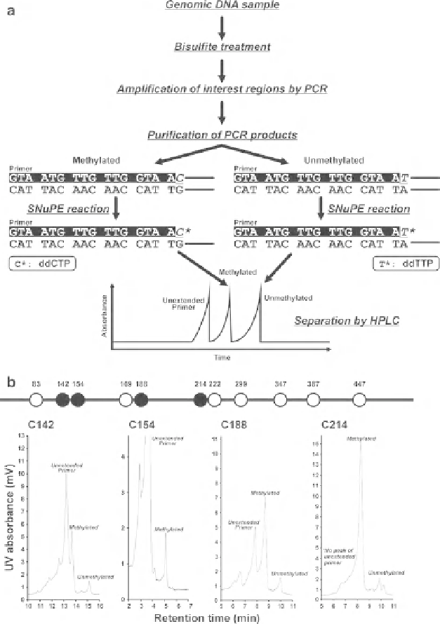
Fig. 10.5
(
a
) Principle of SNuPE analysis. (
b
) Measurement of methylation level of a promoter
region by SNuPE analysis. UV spectra of SNuPE assays analyzing the methylation levels of cyto-
sine 142 (C142), 154 (C154), 188 (C188), and 214(C214). Values in traces show the HPLC reten-
tion times of peaks
the top strand, ddGTP and ddATP when SNuPE primers are placed on the bottom
strand) (Fig.
10.5a
). Primers for an SNuPE reaction should be between 12 and 18
nucleotides long and are designed to match the site immediately adjacent to the
cytosine residue of interest. Taken the T-rich top strand, ddCTP and ddTTP were