Biomedical Engineering Reference
In-Depth Information
9.8
Signal Detection of S
35
-RNA Probe: Visualization
of Radioactive Signal
Detection of Signal with X-ray Film
:
1. Place dry glass slides into a fi lm cassette and expose the slides to X-ray fi lm
(Kodak, BioMax MR fi lm) in a dark room for several days {
Note*1
} (Fig.
9.6a
).
Make sure that the slides face the emulsion side of the X-ray fi lm {
Note*2
}.
2. Develop the X-ray fi lm in standard developer and fi xer in a dark room {
Note*3
}
(Fig.
9.6b
).
3. The hybridization signal shows up as black (exposed silver grains in the emul-
sion) on the fi lm.
Note
*1: For test exposure, usually 1-2 days is enough. For regular exposure, 2-4 days
should be fi ne.
*2: We do not recommend using regular X-ray fi lm that has emulsion on both
sides, because S
35
radioactivity cannot go through opposite side of the X-ray
fi lm. Therefore, regular X-ray fi lms cannot be enhanced with S
35
in situ
hybridization; rather, they cause a more diffused image.
*3: If needed to perform manual development of X-ray fi lm, prepare one developer
container, two tap water containers, and one fi xer container in a dark room.
Under safelight conditions, put X-ray fi lms in a developer container for 3 min,
then transfer it to the fi rst tap water container for 1 min to rinse out developer solu-
tion. Immerse fi lms in a fi xer container for 3 min, transfer it to the second tap water
container for a few minutes rinsing very well, and then hang dry them (the later step
can be at regular room light).
(Optional)
Detection of Signal with High Resolution by Silver Grain Dipping
:
For Generating of Higher Resolution Signals with Cresyl Violet {
Note *1
}:
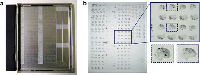
Fig. 9.6
Visualization of radioactive signal with X-ray fi lm. (
a
) Setting of hybridized glasses on a
fi lm casette. (
b
) Developed X-ray fi lm. Black color represents mRNA signals