Biomedical Engineering Reference
In-Depth Information
a
eYFP/
eCFP
6
eYFP
2
4
0
2
-2
eCFP
0
1 s
1 s
Odor
Odor
-4
b
eYFP
4
2
eYFP/
eCFP
2
0
eCFP
0
-2
5 s
5 s
Sound
Sound
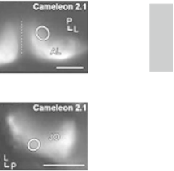
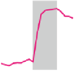
Fig. 7.2
Stimulus-evoked calcium signals. Ca
2+
signals in the AL (
a
) and the JO neurons (
b
) in
response to odor and sound, respectively, were recorded. The
circles
in the
left panels
indicate the
regions in which calcium signals were monitored.
Dashed line
indicates the midline of the brain.
Each stimulus-evoked reciprocal fl uorescent changes (
Δ
F
/
F
0
) between eCFP (
blue line
) and
eYFP (
yellow line
) by FRET (
middle panels
).
R
/
R
0
(%) is the change in eYFP/eCFP fl uores-
cence ratio, where
R
is the average eYFP/eCFP ratio before stimulus onset and
∆
∆
R
is the deviation
from
R
(
right panels
). Scale bar in the
left panels
= 50
m. Lines with characters L and P in the
left panels indicate the directions to the lateral and posterior, respectively [modifi ed from
Kamikouchi and Ito (
2007
) ]
μ
because of wavelength ratioing. A drawback of the ratiometric indicators relies on
the relatively small fl uorescence changes caused by FRET, which results in a lower
signal intensity when compared to single-chromophore sensors. Calmodulin-based
two-chromophore indicators, such as Cameleon (Fiala et al.
2002
; Miyawaki et al.
1999
) and D3cpVenus (Hendel et al.
2008
; Palmer et al.
2006
), or the troponin
C-based indicator such as TN-XXL (Mank et al.
2008
) has been successfully used
in
Drosophila
. Technically, two-chromophore sensors are more complicated to use
as its application requires the simultaneous recording of two wavelengths. A CCD
camera with a beam splitter device or two detectors with different emission fi lters
enables such dual-channel imaging.
By choosing GAL4 fl y strains from the available collections, GECIs can be tar-
geted to virtually any neurons of interest (Fig.
7.2
). These probes translate a change
in ion concentration into changes in the fl uorescence (Palmer and Tsien
2006
).
Thereby, the activity of large populations of genetically and functionally related
neurons can be monitored simultaneously, which overcomes the spatial limitations
of electrode recordings. Whereas electrophysiological recordings using electrodes
monitor changes in membrane or fi eld potential of individual neurons or neuronal
populations, Ca
2+
sensors exploit the fact that changes in membrane potential are