Information Technology Reference
In-Depth Information
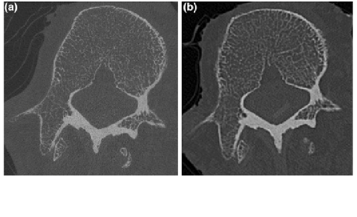
Fig. 9 Corresponding hr-pQCT (a) and MDCT (b) image of a formalin-
xed spinal segment unit.
Spatial resolution was 41
ʼ
m
at hr-pQCT and 250
×
250
×
600
ʼ
m
at MDCT. Note the better
depiction of the single trabeculae in the hr-pQCT image
Standard parameters for the assessment of trabecular bone microstructure can be
calculated in the binarized MDCT images according to bone histomorphometry
using the mean intercept length method [
77
]: Bone volume divided by total volume
(BV/TV; bone volume fraction; [%]), trabecular number (TbN; [mm
−
1
]), trabecular
separation (TbSp; [mm]), and trabecular thickness (TbTh; [mm]). In contrast to hr-
pQCT and
CT, MDCT derived parameters are labeled as apparent values, since
they cannot depict the true trabecular microstructure due to the limited spatial
resolution. Furthermore, several advanced measures of trabecular bone micro-
structure have been introduced, e.g. non-linear topological parameters such as the
Minkowski Functionals [
78
]. The appropriate de
μ
nition of thresholds for image
binarization is critical for the calculation of these trabecular bone microstructure
parameters. The absolute values of these parameters vary with different selected
thresholds due to partial-volume effects. An optimized, global threshold is usually
chosen for MDCT images, so that subjects with dense trabecular bone micro-
structure do not have only bone voxels and osteoporotic subjects not only marrow
voxels. To give an example, Baum et al. [
69
] applied an optimized, global threshold
of 200 mg hydroxyapatite/cm
3
on vertebral bone specimens. To avoid the depen-
dence of the results on the selected threshold, trabecular bone microstructure
parameters have been introduced which do not require a threshold, e.g. the scaling
index method [
78
,
79
]. It reveals the local dimensionality of each voxel (i.e. more
plate-like or rod-like structure). Thus, the transformation of trabecular bone from
plate- to rod-like structures due to osteoporosis can be identi
ed (Fig.
10
). Elastic
and shear moduli obtained from FEMs represent an alternative to the trabecular
bone microstructure parameters to predict bone strength. FEMs can be calculated
not only in ROIs in the trabecular bone, but also integrally for the whole vertebra
including the cortical bone. This is advantageous, since it is well known that the