Information Technology Reference
In-Depth Information
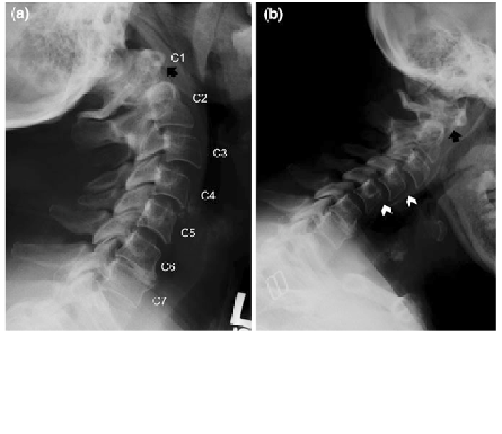
Fig. 12 Plain radiographs of the cervical spine showing atlanto-axial subluxation and subaxial
subluxation in a patient with rheumatoid arthritis. The lateral views of the cervical spine were
taken with neck extension (a) and
flexion position, a separation of anterior arch
of C1 from the dens of C2 (arrow) is revealed. This is not observed in the extension position
(arrow), indicating the presence of dynamic atlantoaxial subluxation. Subaxial subluxation
(arrowheads) is also present at C3
fl
flexion (b). In the
fl
C4 and C4
C5
-
-
fluid related to
RA, as well as bone erosions. In addition, bone marrow edema on MRI may
indicate in
MRI can also demonstrate synovial thickening and excess joint
fl
ammation not otherwise appreciated. However because of its expense
and the fact that most information can be obtained by other modalities, MRI is not
often used in clinical practice for imaging the peripheral joints in RA. However,
MRI is valuable in imaging the cervical spine, particularly in detailing areas of
spinal cord or nerve root compression. Computed tomography is not often used,
because the necessary structural information can in most instances be obtained by
radiography or MRI.
fl
3.6 Juvenile Idiopathic Arthritis
Juvenile idiopathic arthritis is the most common type of arthritis in children, but is
rare, affecting 10,000
60,000 children in the United States. Among several sub-
types, three are most common: pauci-articular, polyarticular, and systemic-onset
subtypes. The pauci-articular subtype, which typically affects girls under the age of
5, presents with in
-