Information Technology Reference
In-Depth Information
3.1.4 Imaging
Plain radiography is important in the diagnosis of AS and in the exclusion of other
diagnoses, particularly in patients with advanced disease. Usually an anteroposte-
rior (AP) view of the pelvis is obtained for evaluation of the structural changes of
the sacroiliac joints. Erosions, sclerosis and ankylosis of the sacroiliac joints are the
common
4, from normal to the most
advanced disease (Fig.
8
and Table
2
). Presence of bilateral grade 2 changes or
unilateral grade 3 or 4 changes is required for classifying AS by the modi
findings in AS. These changes are graded as 0
-
ed
New York criteria. Structures other than the sacroiliac joints can be assessed by
pelvis X-ray. Erosions and loss of the joint space of the hips, and calci
cation along
the tendon insertions are seen in patients with AS.
However, pelvis radiographs have limitations. They have low sensitivity and
speci
am-
mation in the bone or joints. In patients with a short duration of symptoms, MRI of
the pelvis and the lumbar spine is often used to detect early disease. Active
city for bony changes early in the course of AS, and cannot show in
fl
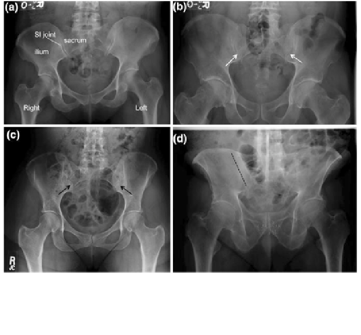
Fig. 8 Radiographic grading of sacroiliac joint involvement in ankylosing spondylitis. a Right
sacroiliac (SI) joint grade 0 (normal); left sacroiliac joint grade 1 (suspicious for changes). b Right
sacroiliac joint grade 2, left sacroiliac joint grade 2 (small localized narrowing, indicated by white
arrows). c Right sacroiliac joint grade 3, left sacroiliac joint grade 3 [partially fused with residual
joint space (black arrows)]. d Right sacroiliac joint grade 4, left sacroiliac joint grade 4 (complete
fusion). Dotted line indicates the location of the fused right sacroiliac joint