Information Technology Reference
In-Depth Information
lead to permanent damage of the nerve, and lead to irreversible nerve symptoms. If
compression of the spinal cord is present with clinical symptoms of myelopathy,
de
nitive surgery is indicated.
2.2.4 Imaging
Plain radiographs of the spine have limited use in diagnosing degenerative disk
disease, although some radiographic
findings indicate degenerative disk disease.
These
findings include narrowing or loss of the disk height (Fig.
4
), sclerotic
changes of the vertebral endplates, and, in later stages, the presence of osteophytes
and sclerosis of the facet joints.
c
radiographic indicator of disk degeneration. With degeneration of the disk, gases
transpired from the circulation accumulate in the space that the nucleus pulposus
once occupied, causing the intervertebral disk space to appear radiolucent (Fig.
4
).
In general, in the absence of trauma, radiographs are not always needed.
“
Vacuum phenomenon
”
is considered a speci
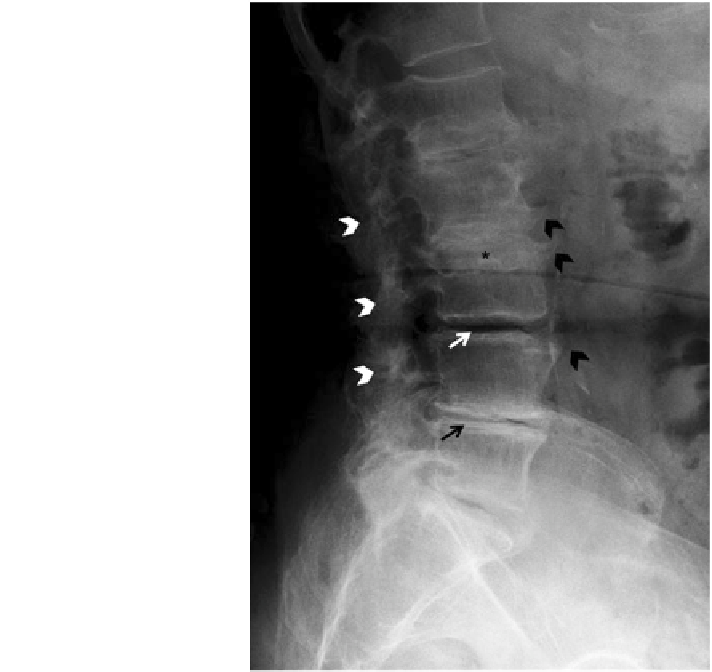
Fig. 4 Severe degenerative
disk disease and lumbar
spondylosis on plain
radiograph. Lateral view of a
lumbar spine. Narrowing of
intervertebral disk space
(black arrow), complete loss
of disk space (asterisk), and
vacuum phenomenon (white
arrow) are characteristic
features of degenerative disk
disease. Osteophytes
originating from vertebral
bodies (black arrowheads)
and facet joint narrowing
(white arrowheads) are
present, signifying associated
spondylosis