Information Technology Reference
In-Depth Information
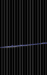
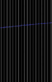
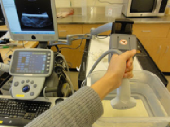
Fig. 8 Moving the transducer up/down repeatedly for acquiring tracking and imaging data for
temporal calibration (left). Position of the water tank bottom is automatically detected in the
ultrasound image and used as position signal for the image data. Position of the water tank bottom
is shown for the top and bottom positions (center, right)
synchronization is not available but the acquisition rate and latency is constant in
both the imaging and tracking device then software-based method can be used to
compute the
fixed time offset. Methods based on detecting certain events (such as
sudden motion) have been proposed. These methods are easy to implement, but
inaccurate or require lengthy data acquisition, because acquisition of a single
measurement sample takes a few seconds. Correlation-based methods require the
operator to perform quasi-periodic motion with the transducer for a few seconds
and during this time imaging and tracking data is recorded (Fig.
8
). Then position
signal is extracted from the data and the time offset is computed that results in the
highest correlation value between the position signals (Fig.
9
). Position signal from
the 3D pose information can be computed as position along the
first principal axis
of the motion. Position signal from the image data can be retrieved by detecting the
position of a feature (such as the bottom of the water tank) and use the position
along a chosen axis. The correlation-based temporal calibration method is accurate,
reliable, and a free, open-source implementation is available in the Plus toolkit [
3
].
7 Volume Reconstruction of Tracked Ultrasound
Position of recorded ultrasound images can be used to reconstruct 3-dimensional
ultrasound volumes. Reconstructed volume data can be in the same format as other
volumetric images (CT or MRI), but the intensity values of voxels still highly
depend on the direction of sound propagation. Therefore, processing and visuali-
zation of such volumetric images are dif
cult. Intensity values in ultrasound are not
characteristic to tissue types, and are often attributed to artifacts (including scatter
and shadow), rather than anatomical structures. Image quality and parameters also
depend on the settings of the ultrasound scanner, the size of the patient, and motion
patterns of the transducer during image recording.