Information Technology Reference
In-Depth Information
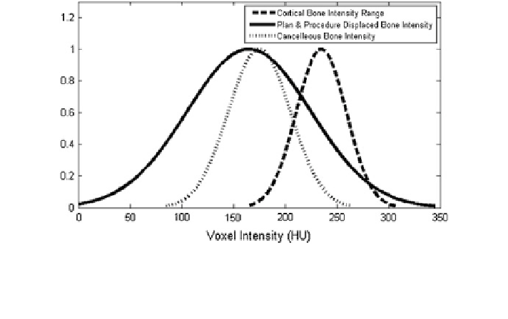
Fig. 12 Voxel intensity distribution for the cancellous and cortical bone according to the linear
relationship between bone mineral density and image intensity. Superimposed voxel intensity
range of the bone volume displaced by the implanted pedicel crews and computed according to the
Fastening Strength formulation
introduced by the surgeon
s skill level and screw measurement
variability, provide similar Fastening Strength to the actual procedures and the
proposed Fastening Strength correlates positively with implant dimension.
'
s versus fellow
'
5 Current Clinical Experience and Relevant Cases
Given the perceived bene
t of the 3D planning method, seven Mayo Clinic
orthopedic surgeons have used the 3D templating tools to pre-operatively plan
several cases.
Case Study 1: A 4 year old male presented with progressive congenital scoliosis
associated with VATER syndrome and neuro
bromatosis. The scoliosis was
present at two levels. The patient had a complex cervicothoracic curve which
progressed from 25
over one year. Hemivertebrae also caused thoraco-
lumbar scoliosis which progressed from 25
°
to 30
°
°
to 35
°
over a one year period, with a
focal kyphosis measure of 22
. For this patient, pre-operative CT scans were
ordered to more precisely determine the pathologic anatomy and to permit 3D
templating. The CT images clearly illustrated pathologic congenital anatomy of
cervical and thorocolumbar congenital scoliosis, as shown in Fig.
13
. 3D templating
con
°
rmed that the thorocolumbar pedicles could safely accommodate 3.5 and 4.0
diameter screws, also shown in Fig.
13
, but the cervicothoracic vertebrae would not
safely accommodate standard implants. The 3D templates allowed for straightfor-
ward generation of a physical model using a commercial 3D printer. This model
was used intra-operatively, showing the precise starting points and trajectory for