Information Technology Reference
In-Depth Information
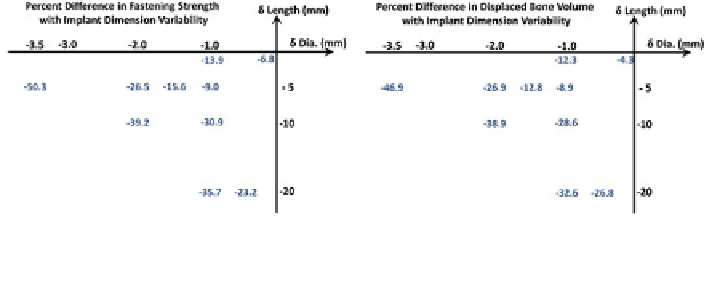
Fig. 11 Percent difference in Fastening Strength and displaced bone volume with variations in
implant dimension. The negative values indicate loss of holding power and underestimated
displaced bone volume due to undersized implants
fastening strength. While the difference in displaced bone volume measurements for
identical size implants are mainly arti
cial, primarily due to partial volume effects
inherent to the CT image resolution, the remaining differences in Fastening Strength
may be real and mainly due to the difference in voxel intensity within the displaced
bone volume.
With regards to the mean voxel intensity of the displaced bone volume, the plan
and procedure both have comparable ranges, and consistent with image intensity
range of the cancellous bone. Recall that cortical spine BMD was estimated as
192
140 mg/cm
3
. Given the
linear relationship between BMD and image intensity, cortical bone features
a
10 mg/cm
3
, while cancellous bone BMD averaged
±
*
235
±
14 intensity range, while cancellous bone averages a mean voxel
*
intensity of
175. As shown in Table
1
, the mean voxel intensity of the displaced
bone volume in both the plan and procedure was on the order of 165
*
60, which
spans the mean voxel intensity of cancellous bone, but also extends into voxel
intensity range associated with the cortical bone (
±
200). From a physical inter-
pretation view point, the screw shaft is immersed into the cancellous bone while the
“
*
”
extends into the cortical bone located toward the edge of the
pedicle body (Fig.
12
), therefore providing added strength that a thinner implant
would not, since only spanning the cancellous bone region.
As documented [
25
,
26
] and also suggested by the orthopedic surgery team, a
pedicle screw implant is typically deemed optimal if the screw fully
tip of the thread
“
taps
”
into the
cancellous pedicle region and the edge of the screw thread
into the hard
cortical pedicle shell for improved holding power [
27
]. Based on the mean voxel
intensity measurements of the displaced bone segment (in both the plans and post-
operative assessments), the screws fully
“
digs
”
“
tapped
”
into the cancellous bone, and also
“
onto the cortical bone, documented by the upper tail of the voxel
intensity range of the measured displaced bone volume (Fig.
12
), con
grabbed
”
rming
clinical requirement. Therefore, our proposed formulation of the Fastening Strength
metric can provide quantitative information whether this clinical requirement has
been met. This demonstrates that
the plans, within their inherent
limitations