Information Technology Reference
In-Depth Information
Typical datasets consist of isotropic images with a 0.75
0.75 mm in-plane
resolution and a 0.75 mm slice thickness. The pre-operative scan is imported into
the SSP software [
14
], within which the surgeon
×
places the pedicle
screws into the 3D image data, generating a virtual surgical plan which can be
loaded up for visualization during the intervention. Moreover, the resulting surgical
plan and image dataset can be further used to generate an appropriate anatomical
model for 3D printing, resulting in a physical, 3D patient-speci
“
virtually
”
c model of the
spine that can be used as a visual aid before and during the procedure.
3.2.2 Patient-Speci
c Virtual Templating
The 3D templating process is the repeated application of two steps for each vertebra
of interest. First, to effectively plan spine surgery using 3D templating tools, it is
necessary to reorient each vertebral body so the axial image plane runs perpen-
dicular to its central axis. To accomplish this task, the user simply identi
es a
bounding box for each vertebra, using the sagittal and coronal views. The top and
bottom sides of the bounding box are aligned with the vertebral endplates, making
sure they extend far enough to include the entire vertebral body and any part of the
implants that will extend outside the vertebra (e.g., the screw heads). This can
quickly be carried out with placement and manipulation of a simple GUI tool with
2
3 mouse clicks in each of the two views, as illustrated in Fig.
2
. The pedicle
lengths and angles are determined in the local vertebral space to ensure that the
measurements correctly represent the anatomy. Also during the vertebral identifi-
-
-
cation process, it may be necessary to reorient each vertebral sub-volume into a
consistent frame of references. Speci
cally, a rotation of the axial image may be
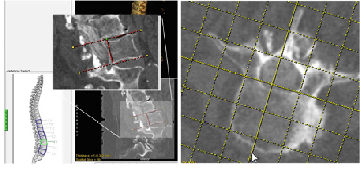
Fig. 2 Vertebral body extraction and alignment. During the process of vertebral body extraction, a
user manually places I-beams around the vertebra of interest (left). After the vertebra has been
extracted, it can be reoriented along the spinous process using an interactive grid (right)