Information Technology Reference
In-Depth Information
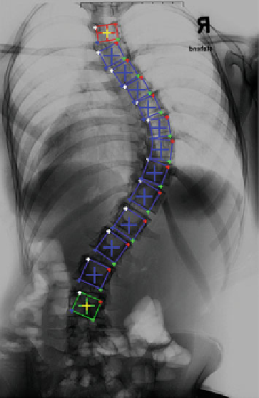
Fig. 5 Results of automatic
vertebra detection from a
low-dose X-ray radiograph of
a scoliotic patient. All 13
visible vertebrae have been
successfully detected by our
algorithm
For the low-dose X-ray radiography of the scoliotic patient, our algorithm can
successfully detect all 13 vertebrae. Figure
5
shows the detection results.
4 Discussions and Conclusions
In this chapter, different from previous work [
9
,
10
], we proposed a graphical
model-based method for automated detection of vertebral bodies from X-ray image
(s). We validated our method on DRRs of twenty-one cadaver spinal segments of
different regions as well as on one low-dose X-ray radiography of a scoliotic
patient. Compared to previously introduced approach, our approach has following
advantages: (1) it does not need to be trained using training data, (2) it does not ask
for the prior information of the examined anatomical region and (3) it can auto-
matically identify the number of vertebrae visible in the image(s) and therefore does
not ask for a prior information about the number of vertebrae to be identi
ed. Our
future work focuses on investigating the performance of the proposed approach on
more clinical X-ray images.
Acknowledgments The authors gratefully acknowledge the
financial support from the Swiss
National Science Foundation through the National Centers of Competence in Research CO-ME.