Information Technology Reference
In-Depth Information
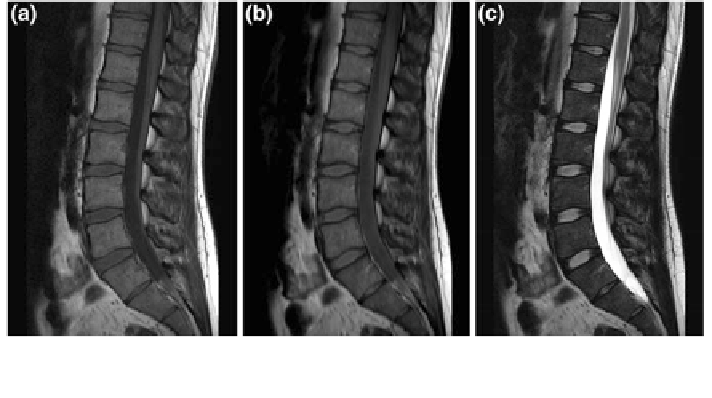
Fig. 12 Spine protocol images in varying sequence weightings. Sagittal images of the lumbar
spine imaging sequences demonstrating a T1, b PD, and c T2 imaging
(black on the display monitor). On T1 imaging,
fluid in the tissues presents as
intermediate to low signal intensity, and fat as high signal intensity. In opposition,
fl
fl
fluid on T2 appears as high signal and fat as high signal. Thus, in principle, one
could differentiate
fluid on a T2 sequence by comparison with a T1 sequence (see
Fig.
12
). There are exceptions to these signal intensity norms, including proteina-
ceous
fl
fluid in the body which can appear high signal intensity on T1.
Modi
fl
cations of these basic sequences have been devised to expand their range
of clinical utility, including what are termed fat suppression variations. In fat
suppression, a process is applied by which adipose tissue, normally high signal
intensity on T1 and T2, is turned to low signal intensity. One method by which this
is accomplished is called fat saturation. Fat saturation depends on the slight
field-
dependent variance in precession frequency between the protons of fat and water
and uses a frequency selective applied excitation to nullify the fat signal intensity
(Fig.
13
)[
8
,
9
]. Consider an example of the clinical usage of fat saturation on a
standard T2 sequence which demonstrates high signal intensity for both fat and
fl
fluid. If the fat signal intensity is now turned low after the application of fat
saturation,
fluid will now be the principle residual high intensity entity left on the
image, amidst an intrinsically dark background of surrounding tissues composed of
shades ranging from gray to black. Thus, any structure with
fl
fluid or edema (of
interest in detecting pathology) will appear prominent, facilitating the detection and
characterization of pathological tissue.
However, fat saturation does not always saturate the images with spatial uni-
formly. The effect of fat saturation depends on the resonant frequency difference
between water and fat, and is subject to variable inactivation when non-uniformities
occur in the applied magnetic
fl
field. This inactivation may occur locally when the
patient has had metallic hardware placed, as in the case of spinal
fixation hardware.
Variable inactivation may also occur near the margins of the magnet or body part