Information Technology Reference
In-Depth Information
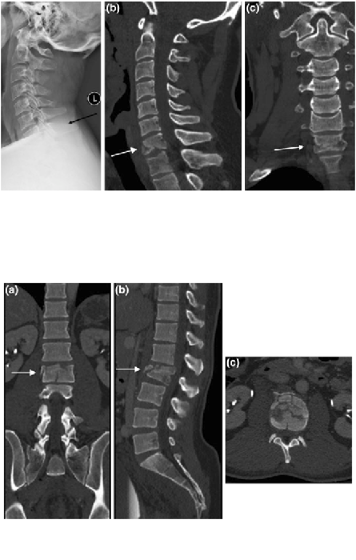
Fig. 9 Diagnostic advantage of CT over radiographs. Patient with history of trauma and neck
pain. In the lateral view radiograph of the cervical spine (a), patient
is injury is not well seen
(at level of black arrow). Sagittal (b) and coronal (c) reformatted images from patient
'
'
s CT scan
demonstrates comminuted fracturing of the C7 vertebra (white arrows)
Fig. 10 Spine protocol CT images. Multiplanar reformatted images from a spine protocol CT
scan, in a sagittal, b colonal, and c axial planes, facilitate the extraction of detailed information
regarding the vertebral fracture pattern and extent allowing a more accurate assessment for
treatment planning