Information Technology Reference
In-Depth Information
1 Introduction
Localization, labeling, and segmentation of the vertebrae and the intervertebral
discs are essential tasks that have been attracting an increasing number of research
groups worldwide. The accuracy and robustness of these imaging tasks are crucial
for subsequent abnormality diagnosis. Moreover, accurate results of these tasks are
critical for radiologists to perform an accurate diagnosis from various imaging
modalities including X-ray radiography, Computed Tomography (CT) scans, and
Magnetic Resonance Imaging (MRI). Furthermore, surgeons demand accurate
reporting of these results when overlaid on a computer guided surgery system or a
computer assisted surgery system.
Whilst the localization task is to locate an anatomical structure (e.g. locating the
intervertebral discs by a point within or a bounding box around the discs), the
segmentation task is to provide a
fine contour that accurately delineates that
structure (e.g. a contour around the vertebra). Labeling, on the other hand, is to
identify the anatomical nomenclature of each structure (e.g. labeling each of the
ve
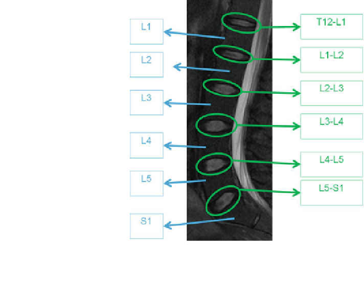
lumbar vertebrae as L1, L2, L3, L4 and L5). Figure
1
shows an example of
localization and labeling for the six intervertebral discs connected to the
five lumbar
vertebrae on a sagittal MRI [
5
].
Fig. 1 Localization and
labeling of a sagittal lumbar
T2-weighted MRI. Lumbar
area is the second area to the
last of the vertebral column. It
is the main part of the
vertebral column that is
responsible for bearing the
major body weight. The
lowest lumbar vertebra is L5
and the highest is L1. Inter-
vertebral discs are labeled
based on the enclosing
vertebrae [
5
]