Information Technology Reference
In-Depth Information
2 Evolution of 3D Reconstruction Systems
2.1 First Generation
For years, research in the
field of biomechanics has focused tremendously on the
analysis of spinal deformities in 3D. The elaboration of complex biomechanical
models required rigorous clinical experimentations, as well as quantitative evalua-
tions of the patient
s posture and movement analysis in three dimensions. In 1971,
Panjabi and White illustrated the importance of studying the spine in three dimen-
sions (3D), with 6 degrees of freedom. A number of techniques for 3D measure-
ments, imaging and modelling of the spine were developed [
1
,
9
,
11
,
24
,
34
]. Since
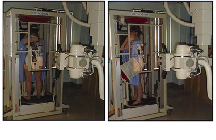
1992, an imaging system installed at the Sainte-Justine Hospital Research Center
enabled to perform the 3D reconstruction of bony structures from radiographic
images (Fig.
2
). The proposed system was based on the calibration principle of the
Direct Linear Transform (DLT), which implicitly includes the geometric parameters
in the coef
'
cients of the projection matrices obtained linearly by an inversion of the
matrix. Using a Plexiglass cage with encrusted steel pellets, the 2D/3D con
guration
could be determined in a linear fashion. This tool was used in over 6,000 patient
visits for research purposes, in addition to other applications such as surgical sim-
ulations and biomechanical analysis of the spine. The system was also used to
improve the quality of diagnosis and follow-up exams for patients with idiopathic
scoliosis. Finally, this 3D imaging system was used in a number of other projects
involving computer assisted surgery and personalization of models.
Fig. 2 One of the first 3D reconstruction systems installed at the Sainte-Justine Hospital in 1992,
where a plexiglass cage with embedded pellets is used to calibrate the X-ray scene. The patient is
turned from the frontal to sagittal position to obtain biplanar radiographs