Information Technology Reference
In-Depth Information
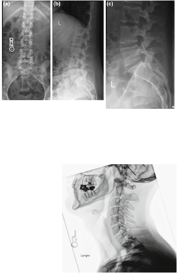
Fig. 1 Illustration of beamline passage through multiple tissue layers and resultant superposed
anatomic structures. Images from a radiographic series of the lumbar spine a frontal view, b lateral
view, c coned down lateral view. Bowel loops most prominently over-project the spine on the
frontal view on the image, due to photon passage through gas
filled hollow viscera. Lateral and
coned down lateral views demonstrate photon passage through (at different levels) and over-
projection of the spine by the lungs, bowel loops, diaphragm, and internal organs (liver and spleen)
Fig. 2 Tissue density
variation as manifested on
radiographs. Lateral cervical
spine radiograph of a 25 year
old male, demonstrating
examples of pixel intensity
correlation with tissue
density, or X-ray beam
attenuation. From lesser to
greater density: a air, b fatty
tissue, c muscle, d bone,
e metallic dental hardware
d
e
c
b
a