Information Technology Reference
In-Depth Information
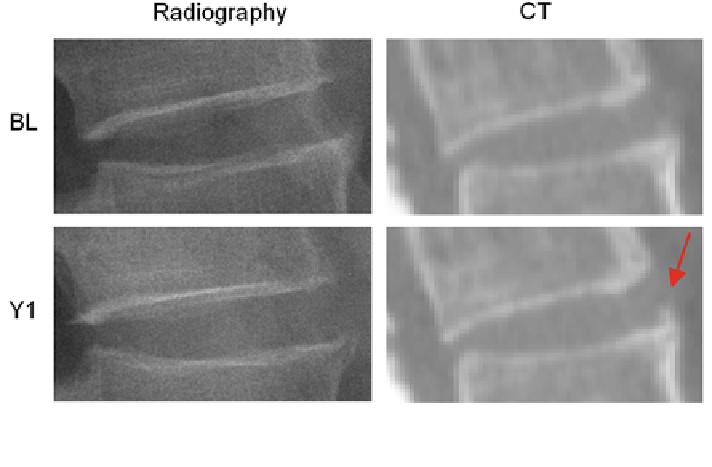
Fig. 1 Example of syndesmophyte growth from baseline (BL) to year 1 (Y1) visible on CT
reformations but not on radiographs
2 The Algorithm
The complete algorithm, summarized in Fig.
2
, has of three main parts. First,
vertebral bodies are segmented using a 3D multi-stage level set method. Triangular
meshes representing the surfaces of the segmentations are made [
34
]. The 3D
surfaces shown in Fig.
2
are triangular meshes obtained from our segmentation
results. The vertebral surfaces of corresponding vertebrae are then registered. The
purpose of the registration is to extract the syndesmophytes of both vertebrae using
the same reference level. Syndesmophytes are cut from the vertebral body using the
end plate
'
s ridgeline as the reference level.
2.1 Segmentation of the Vertebral Bodies
Many image processing segmentation techniques have previously been applied to
the extraction of vertebral bodies in CT [
35
-
42
]. For our algorithm, we chose to use
level sets for their
flexibility [
43
]. Flexibility is essential in our application as
syndesmophytes can deform the normal vertebral shape in unexpected ways. Level
sets are evolving contours or surfaces that can expand, contract, and even split or
merge. For the purpose of segmentation they are designed to deform so as to match
an object of interest. Many different types of level set exist, depending on the image
features chosen to guide the segmentation. For our particular purpose, we selected
fl