Information Technology Reference
In-Depth Information
The results of the previous steps are used to determine the initial location and size
of the parts in the following steps. In our current model, we de
ne 36 border atoms
for the disk model and 20 atoms for the slab models.
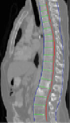
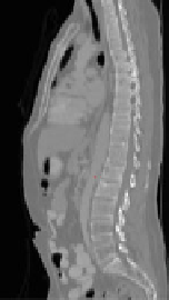
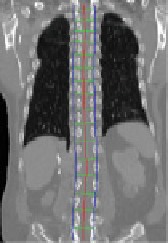
5 Spinal Column Partitioning
The spinal column consists of a set of vertebrae separated by inter-vertebral discs
(Fig.
5
a). Since the spinal column is a curved structure, the standard planar
reformations (sagittal and coronal) do not provide clear views of the vertebral
separation (Fig.
5
b). Curved planar reformation (CPR) (Fig.
5
c) [
42
] is generally
considered superior.
After the spinal column is segmented, we need to partition the spinal column into
vertebrae at the inter-vertebral disc locations so that we can process the vertebrae
separately and also localize the abnormality at the vertebra level. We developed a
partitioning approach based on curved reformation along the spinal canal.
The centerline of the spinal canal is used as the central axis for the CPR. We
generate the CPR in sagittal and coronal directions. Given that the vertices on the
(b)
(c)
(a)
(g)
(e)
(f)
Vertebral
disks
(d)
Aggregated Intensity Profile
1500
1400
1300
1200
1100
1000
900
800
1 4 7 10131619222528313437404346495255586164677073767982
Slice
Fig. 5 Spine partitioning. a Spinal column; b regular sagittal reformation; c curved planar
reformation in sagittal direction; d aggregated intensity profile (AIP) along the spinal canal;
e spinal partitioning in sagittal direction; f spine partition in coronal direction; and g spine partition