Biomedical Engineering Reference
In-Depth Information
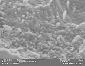
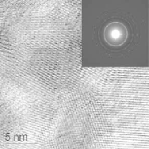
(a)
(b)
(c)
(d)
Figure12.3
FESEMimagesof (a)as-preparedand(b) calcinedTyzor-LA-CNXLcomposite,
andTEMimages(c)correspondingtitaniaproductand(d)high-resolutionTEMimagesof the
titania (inset: selectedelectrondiffractionof titania) (reprintedwithpermission fromref 24).
Copyright2007Elsevier.
and 5-10 nm in diameter. After calcination, it is difficult to discern CNXL-shaped
titania due to thermal collapse. It is apparent that Tyzor-LA-CNXL composites under-
went serious shrinkage during the calcination process. A few aggregated titania showed
CNXL-shapes. TEM images and selected area diffraction pattern (SEAD) were employed
for further determination of the morphology and orientation of the titania. In TEM
images, there are many channels with irregular diameters connected together, which have
been generated by CNXL templates. After thermal treatment, collapsed CNXL-shaped
pores are mesoporous, which are marked with arrows in Figure 12.3c. High resolution
TEM image (Figure 12.3d) shows nanocrystalline anatase lattice. An electron diffraction
pattern recorded on a titania surface has clear diffraction rings that correspond to the
anatase phase of titania. Titania products are clusters of 5-7 nm nanocrystalline anatases.
BET results (not shown here) indicate that titania samples have 170-200 m
2
/g of specific
surface area and irregular pore diameters after calcination at 500
◦
C in air. In addition,
titania samples showed high stability without changing morphology and crystallinity in
harsh media such as conc. HCl, HNO
3
, and dil. H
2
SO
4
. This novel titania material with


Search WWH ::

Custom Search