Biomedical Engineering Reference
In-Depth Information
OH
OH
4
4
3
1
3
1
OH
OH
5
O
5
O
2
2
O
HO
O
HO
O
2
2
5
5
4
3
1
4
3
1
HO
O
HO
O
O
OH
O
OH
OH
OH
Figure10.1
Chemicalstructureofcellulose.
(Figure 10.1) (Fengel and Wegener 1983). The polymer chains are arranged in an
hierarchical order from elementary fibrils of cross dimension 2-5 nm in plant celluloses
(Hon and Shiraishi 1991; Ding and Himmel 2006).
In the plant cell walls, the cellulose microfibrils result from the combined action
of biopolymerization spinning and crystallization. All these events are orchestrated by
specific enzymatic terminal complexes (TC) that act as biological spinnerets. If the TCs
are not perturbed, they can generate endless microfibrils having only a limited number
of defects or amorphous regions (Brown 1996; Brown 2004). These regions are located
on segments of the elementary fibril which are distorted by internal strain in the fiber to
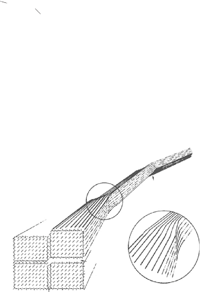
undergo tilt and twist (Figure 10.2) (Rowland and Roberts 1972).
After an acid treatment that hydrolyzes the cellulose and consequently cuts the
microfibrils at each defect, true cellulose rod-like nanocrystals are obtained that have a
morphology and crystallinity similar to the original cellulose fibers. Acids preferentially
C
C
B
C
A
Figure10.2
Schematicrepresentationof theelementaryfibril illustratingthemicrostructure
of the elementary fibril and strain-distorted tilt and twist regions (defects) (Rowland and
Roberts1972, reprintedwithpermissionof JohnWiley&Sons, Inc.).


Search WWH ::

Custom Search