Biomedical Engineering Reference
In-Depth Information
native CNXLs had a better reinforcing effect than the silylated CNXLs and immobilized
the CAB matrix in the vicinity.
A more recent study compared the effect of CNXLs from various sources including BC
to reinforce thermoplastic starch (74). Using different processing methods, thermoplastic
starch and pectin were blended with CNXLs. Namely films were produced by solution
casting with 3% CNXLs while monofilaments were produced by mixing in a Hobart
mixer, followed by extrusion. After equilibration at 50% RH the tensile properties of
the films were determined and the thermal properties of samples with and without CNXLs
were compared. The Young modulus of thermoplastic starch increased from 1.39 GPA in
the neat state to more than 6 GPa with the incorporation of bacterial CNXLs. Elongation
at break also increased from 2.7% to 4% with bacterial CNXLs. While bacterial CNXLs
clearly improved the performance of thermoplastic starch, they were not as effective as
CNXLs from softwood or cotton. For example the elongation at break was 8% with
softwood or cotton CNXLs, that is twice that observed with bacterial CNXLs. Blends of
starch and pectin (50/50) were also produced and exhibited better strength and stiffness
than the pure thermoplastic polymers. However, regardless of the CNXLs origin, their
incorporation in the thermoplastic blend decreased its strength, elongation and tensile
modulus altogether.
In another study, bacterial CNXLs were incorporated in polyethylene(oxide) and effec-
tively electrospun in a 1.5 mm capillary tube (75).
The BC CNXLs had dimensions
of 420
2 nm as measured by TEM and AFM and
when added to PEO as a suspension with some additional water, the PEO/cellulose
whiskers mix was amenable to electrospinning in terms of viscosity and surface tension.
Interestingly the nanofiber diameter increased with the incorporation of the cellulose
nanowhiskers.
±
190 nm X 11
±
4nmX10
±
20 nm which
increased to the 250-350 nm range with incorporation of 0.2 wt% and 0.4 wt% cellulose
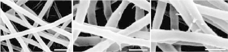
whiskers. The nanofiber size distribution was also increased with the incorporation of
the cellulose whiskers (Figure 9.31). The cellulose whiskers were well incorporated into
the nanofibers, although some whiskers were observed to protrude out of the nanofiber
(Figure 9.32).
For neat PEO the nanofibers had a diameter of 140
±
Further morphological investigation of the cellulose whisker dispersion
0.5
µ
m
0.5
µ
m
0.5
µ
m
(a)
(b)
(c)
Figure 9.31
Field emission scanning electron microscopic (FESEM) image of electrospun
PEO/cellulose whiskers having (a) 0 wt%, (b) 0.2 wt% and (c) 0.4 wt% of whiskers (Park,
W.-I.;Kang,M.;Kim,H.-S.; Jin,H.-K.,Electrospinningofpoly(ethyleneoxide)withbacterial
cellulosewhiskers,Macromolecular Symposia,2007,249-50,289-94.CopyrightWiley-VCH
VerlagGmbH&Co.KGaA.Reproducedwithpermission.)
Search WWH ::

Custom Search