Chemistry Reference
In-Depth Information
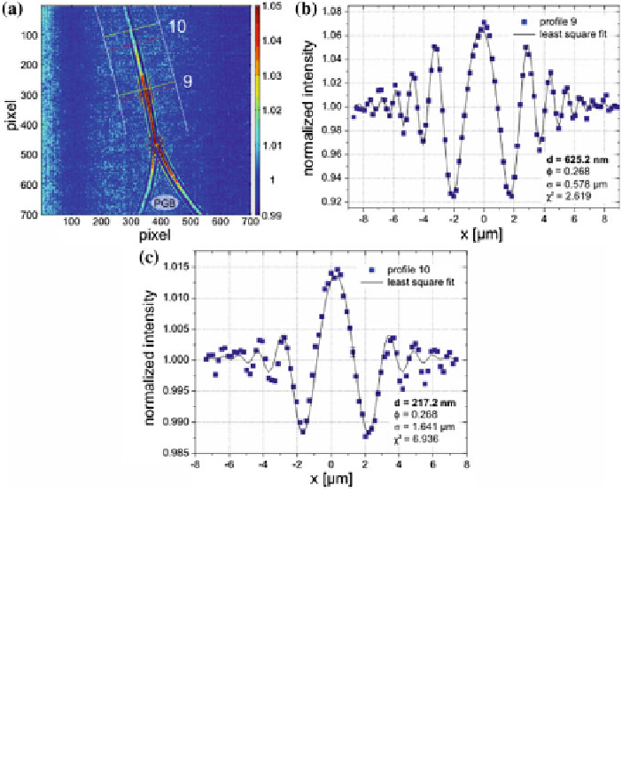
Fig. 4.7 a
Diffraction pattern of the transition region during the “zipper-like” effect of a microflu-
idic lipid membrane close to the Plateau-Gibbs border (PGB) (Monoolein-squalane/water,
z
eff
=
.
94
21mm). The Fresnel fringes disappear in regions where the membrane has already thinned and
only a single maximum is left.
b
Profile 9
, along with a reasonable least-squares fit, showing a
large thickness at the position of a thick, domain of residual solvent.
c
Profile 10
, which is located
close to the bimolecular membrane region, cannot be fitted comparably accurately due to a notable
contribution of the PGB to the diffraction pattern
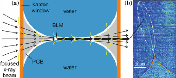
Fig. 4.8 a
Schematic representation of the “focusing” effect of the Plateau-Gibbs border. This
results in an intensity maximum in (
b
) at the position of the thinned membrane. Images of
b
data:
hunt01-7150
, background:
hunt01-10049