Chemistry Reference
In-Depth Information
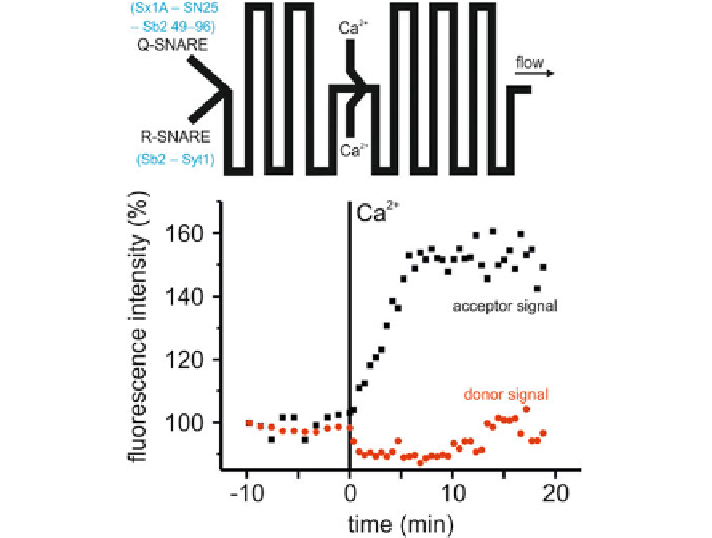
Fig. 3.9
Microfluidic triggering of membrane fusion at low ionic strengths by mediating
Ca
2
+
-
synaptotagmin-1 interactions.
Top panel
The channel geometry to study the
Ca
2
+
triggering. The
Q-SNARE and the R-SNARE are initially mixed and their fusion kinetics can be followed by taking
images at multiple turn segments. After a certain time,
Ca
2
+
is introduced from a side channel from
which point on, the
Ca
2
+
triggered fusion kinetics can be studied.
Bottom panel
The fluorescence
intensities of the donor and acceptor fluorophore are shown as a function of time. The time point
of
Ca
2
+
addition is taken as t
0. Before the addition of
Ca
2
+
no fusion takes place in spite of the
synaptotagmin-1 already present in the R-SNAREs. As soon as
Ca
2
+
is added, fusion increases
dramatically
=
did not progress beyond the hemifusion state. To resolve this issue, we employed a
content mixing assay where liposomes with encapsulated calcein at self quenching
concentrations were fused with empty (calcein-free) liposomes [
25
]. Content mix-
ing results in calcein dequenching. Indeed, a SNARE and
Ca
2
+
-synaptotagmin-1
specific increase in fluorescence was observed (Fig.
3.11
). This content mixing was
not caused by leakage of the calcein from the liposomes, as leakage was only 4-5%
of total calcein.
Synaptotagmin-1 in concord with
Ca
2
+
seemingly overcomes the repulsive inter-
actions to induce membrane fusion at low ionic strengths. What exactly is the
mechanism by which this trigger is mediated? As we discussed earlier, a variety
of mechanisms have been proposed in the past by which synaptotagmin-1 affects
the membrane to trigger fusion. In the next sections, the putative mechanism at low
ionic strengths and the interactions with
Ca
2
+
is investigated.