Chemistry Reference
In-Depth Information
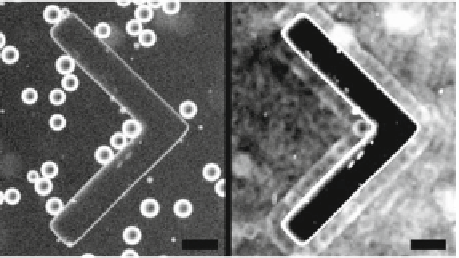
Fig. 7.8
Squirmers at angu-
lar walls.
Left
Squirmers are
observed in a 2 dimensional
space with a V-shape wall
made from PDMS in the mid-
dle.
Right
An overlay of 600
images shows an aggregation
near the wall, in particular as a
monolayer. The contrast of the
image has been enhanced for
improve visualisation.
Scale
bar
100 microns
counted. We therefore find that the droplet squirmer number profile peaks strongly
at the wall, indicative of the attractive interactions with walls. Together with the
previous observation, we conclude that the droplet squirmers aggregate near walls
and swim along them for extended times.
Next, we replace straight walls with angular walls in the middle of a channel
as shown in the left panel of Fig.
7.8
to see if droplets tend to accumulate more
along concave walls than convex ones. The chevron shaped structure has angles of
90
◦
at all edges. The attraction of the swimmers to these walls can be seen in the
right panel of Fig.
7.8
. This image is constructed by overlaying 600 images (each 1
second apart) of the swimming droplets as the one shown in the left panel. While the
moving droplets appear as a dark smear on the image, it can be seen that along all
the walls, the imprints of a monolayer of droplets can be clearly seen, indicating that
they spend a significant amount of time swimming along the walls. Particularly, at
the concave corners, the swimmers are trapped for a very long time. As we will see
below, the trapping due to the asymmetric V shaped geometry, combined with the
swimmer property of travelling along the walls, can be used to rectify the motion of
a population of the swimmers.
In a set of very elegant experiments, Galajda and colleagues [
26
] constructed a
bacterial rectifier using a chamber that was divided into two halves by V-shaped walls
as shown in Fig.
7.9
. When a bacterial population was initially uniformly distributed
in the chamber, the bacteria were found to be concentrated in one of the halves
(right) with the passage of time, as shown in the fluorescence image. This effect
was attributed to the fact that bacteria were self propelled swimmers that can travel
ballistically along walls and a purely physical mechanism was proposed to explain
the phenomenon, not involving any biochemical mechanisms such as chemotaxis. It
was proposed that for a geometry of walls separating two halves of a chamber, as
shown in the bottom panel of Fig.
7.9
, bacteria swimming towards the walls from the
left (numbered 1 and 2) tend to swim along the walls thus leading them into the other
side of the chamber through the gap between walls. However, when they come in
from the right (numbered 3 and 4), due to the shape of the walls, the bacteria which
swim along the walls will be guided back into the same side of the chamber. Thus,
on average more bacteria travel from left to the right than the other way, leading