Biology Reference
In-Depth Information
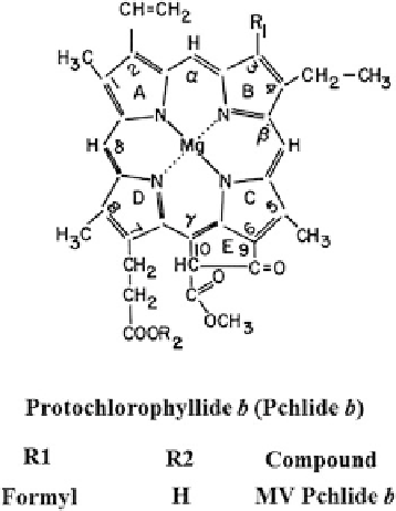
Fig. 12.1 MV Pchlide
b
of a formyl instead of a methyl group at position 3 of the macrocycle 9 (Fig.
12.1
).
The trivial name MV Pchlide
b
was proposed to differentiate it from MV Pchlide
a
.
Determination of the Amount of MV Pchlide
b
either in the Presence of MV Chl
(ide)
a
and
b
, or in the presence of MV Pchlide
a
was achieved by combined
spectrofluorometric analysis at room temperature and 77 K (Ioannides et al.
1997
).
In green cucumber seedlings grown under a 14-h light/10-h dark photoperiod, the
amount of MV Pchlide
b
ranged from about 400 to 800 nmoles per 100 mg proteins.
MV Pchlide
b
was detectable in green tissues but not in etiolated tissues or during
the early phases of greening of etiolated tissues (Kolossov and Rebeiz
2004
).
12.2.1 Arguments Related to the Spectral Properties
of Synthetic Putative Pchlide b
In a recent review Rudiger proposed that the synthetic and natural MV Pchlides
b
describedby(Shedbalkaretal.
1991
), do not correspond to authentic Pchlide
b
(Rudiger
2003
). It was argued that this is because the synthetic MV Pchlide
b
prepared
by Shedbalkar et al., exhibits different absorbance and mass spectroscopic properties
than the putative Pchlide
b
prepared by Schoch et al. (
1995
). While the synthetic MV
Pchlide
b
prepared by Shedbalkar et al., exhibited a typical Pchlide spectrum with band
I and band II maxima at 632 and 582 nm and a band II:I ratio of 0.45 in acetone at
room temperature, Schoch et al. putative Pchlide
b
exhibited a quasi-oxorhodo,
protopheophytin type spectrum with band I and II maxima at 622 and 578 nm
respectively and a band II:I ratio of about 1.57. As a consequence Rudiger goes on
to propose that contrary to the assertions of Shedbalkar et al., MV Pchlide
b
does not
really occur in green plants (Rudiger
2003
).
Search WWH ::

Custom Search