Biomedical Engineering Reference
In-Depth Information
He rce pt o in
Anti He r 2
Erbitux
Anti-He r1
RGD
O
O
O
O
O
O
O
O
O
-O
P
OH
-O
P
OH
-O
P
OH
O
O
O
O
O
O
O
O
O
O
O
O
O
O
O
C13
C12/C13
C1 2
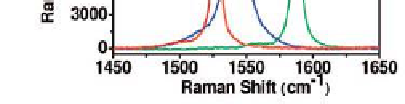
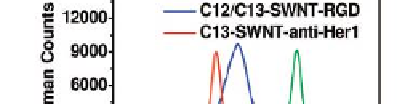
Figure 9.14
SWCNTs with different Raman colours. (a) Schematic of SWCNTs
conjugated with different targeting ligands. (b) Solution-phase Raman spectra of the
three SWCNT conjugates under 785 nm laser excitation. Figure partially modiied
from Liu
et al
.
72
with permission. See also Colour Insert.
The Raman technique was also used to evaluate tumour targeting and
localisation of SWCNTs in living mice, after intravenous injection.
74
Since
Raman imaging was able to detect increased accumulation of targeting
SWCNT-RGD in tumour as opposed to plain SWCNTs (with remarkable
statistical signiicance,
p <
0.05), these results encourage the development of
a new preclinical tool based on the Raman imager.
Another recent development in the ield of diagnostics is represented
by the label-free biosensor obtained from SWCNTs coated with a speciic
rheumatoid arthritis (RA)-peptide and deposited on a quartz crystal
microbalance (QCM) sensing crystal.
75
Results deriving from the detection
of RA antibodies in serum by QCM sensing indicated a higher sensitivity (in
the femtomolar range) of the nanotube-based sensor, in comparison with
both the native peptide and other conventional approaches such as ELISA
and microarray; moreover, the CNT-based device demonstrated a better


Search WWH ::

Custom Search