Biomedical Engineering Reference
In-Depth Information
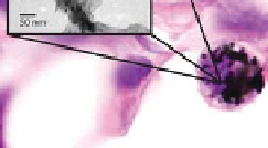
An interesting investigation has been reported on the adverse effects
determined by aerosol release obtained through the mechanical collisions of
as produced SWCNTs and bronze beads.
47
Even though the article provided
additional insights on the toxicity of CNTs, the production of ultra-ine powders
was not very eficient, and it was not possible to discriminate if such particles
were made up mainly of nanotubes or of catalyst. As a consequence, most
probably the respirable fraction of the nanotube aerosol was smaller than the
estimated mass concentrations indicated above. Even more important, in none
of the cases reported there was any indication that handling the nanotube
material led to an increased concentration of ine particles, suggesting that
eventual released particles tended to be larger than 1 μm in diameter. Anyway,
it is worth mentioning that, although nanotubes tended to agglomerate into
nanoropes, thus reducing the formation of an appreciable respirable aerosol
(with estimated airborne concentrations of nanotube material lower than 53
μg/m
3
), it is also possible that such nanoparticles remain in the mouth and
nasopharyngeal regions, still causing a potential health risk.
In the same manuscript, the authors also reported a preliminary
quantiication of the amount of nanoparticles deposited on the gloves of
normal workers: potential dermal loading of SWCNTs was estimated by
placing cotton gloves over the rubber gloves generally used by the workers.
Glove deposits of SWCNTs during handling were estimated at between 0.2
mg and 6 mg per hand. Even though the maximum concentration of 6 mg
is quite alarming, it is very likely that it is overestimated to a certain extent.
In fact the cotton gloves used to collect the hand samples are likely to retain
more material than latex gloves (or similar) or nude skin, so these results
are useful to encourage protective measures, but they also require additional
investigations.
CNTs
macrophage
10 μm
Figure 8.4
Inhaled carbon nanotubes accumulate within cells at the pleural lining
of the lung as visualised by light microscopy. Reproduced from
www.nanotech-now.
com/news_images/35119.jpg with permission of Dr James Bonner, North Carolina
State University. See also Colour Insert.


Search WWH ::

Custom Search