Biomedical Engineering Reference
In-Depth Information
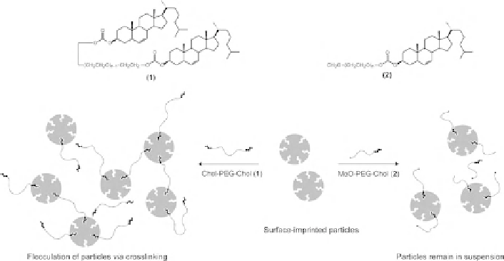
Fig. 7 “Immunoprecipitation-like” separation of surface-imprinted particles in the presence of
(PEG)-bis-cholesterol. The addition of the multi-ligand template resulted in flocculation of MIP
particles (adapted with permission from [
103
])
Another possible strategy to exploit MIP NPs in assays would be to make them
fluorescent [
104
,
105
]. Diltemiz and coauthors grafted CdS quantum dots with a
MIP for guanosine [
38
]. MIP nanoparticles had an average diameter of 45 nm, and
their intrinsic fluorescence was enhanced by binding of the template, proportionally
to its concentration. MIP nanoparticles exhibited a high response to guanine and
guanosine, while adenosine did not give rise to any change in fluorescence. Purely
organic MIP nanoparticles with fluorescent sensing capability were recently
prepared by Ivanova-Miteseva et al. who prepared a fluorescent core by partially
modifying the peripheral amino groups of a poly(amido amine) (PAMAM)
dendrimer with dansyl residues [
106
]. The remaining free amino groups were
then modified with diethyldithiocarbamate (iniferter) groups capable of initiating
photochemical polymerization of an imprinted polymer shell. The particles had an
unusual cube-like shape and were 50 nm in size. The fluorescent MIP NPs (but not
blank NPs) showed an enhancement of fluorescence in the presence of the template
(acetoguanamine) with a detection limit of 30 nM, but did not respond to close
structural analogues. Recently Li and coauthors have developed 350 nm MIP NPs
with a double-layer core-shell structure made of a Fe
3
O
4
nanoparticle core, an inner
shell of fluorescein isothiocyanate and an outer MIP shell, for faster separation and
recognition of E2 [
107
]. The MIP shell was produced using a controlled living
RAFT polymerization. The fluorescent intensity of MIP NPs decreased with
increasing concentrations of E2, showing a detection limit of 0.19
M. They
exhibited a discrete imprinting effect and very good selectivity. Such a system
not only provided a source of fluorescence but also allowed magnetic separation to
replace centrifugation and filtration steps during the experimental procedure.
m
Search WWH ::

Custom Search