Biomedical Engineering Reference
In-Depth Information
2.3
Electron probe in imaging technology.
However, a full quantitative understanding of the CMR effect remains
elusive and it is still the subject of significant research.
In trying to understand the mechanism behind CMR, scientists have used
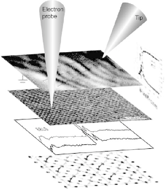
an electron probe to make images and collect other data while using a
scanning tunneling microscope tip to apply current or an electric field to the
sample. In Fig. 2.3, the first layered image of black lines shows polaron
waves, which propagate during the application of the current. Fine dots in
the second layer are the individual atoms, while the periodic dot-clusters
show the electron ordered state. The graph of electron energy loss
spectroscopy (EELS) reveals bonding-electron excitation. The bottom
layer is a structural model of the crystal lattice and the vertical graph
shows the electric resistance (I-V curve) of the crystal when current is
applied.
Experiments at the U.S. Department of Energy's Brookhaven National
Laboratory have shed new light on some materials' ability to dramatically
change their electrical resistance in the presence of an external magnetic or
electric field [139-141]. The Brookhaven scientists studied crystalline
perovskite manganites that had been doped with extra charge carriers -
electrons or 'holes' (the absence of electrons) - using various state-of-the-art