Biomedical Engineering Reference
In-Depth Information
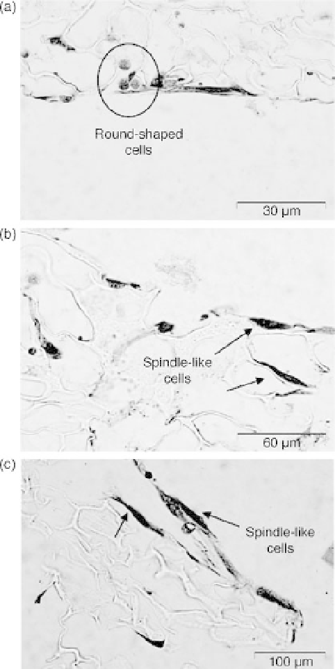
17.8 Optical microscopy photographs of colored MSCs (H&E staining)
attached to (a) neat PLLA, (b) PLLA/mHAP and (c) PLLA/nHAP. Reprinted
with permission from Elsevier (Nejati et al., 2008).
noticeable on the surface of pure PLLA scaffold (Fig. 17.8(a)) while
proliferated cells on the micro- and nanocomposite scaffolds exhibit spindle-
shaped morphology (Fig. 17.8(b) and (c)). The PLLA/HA scaffolds
appeared to be in-vitro biocompatible and non-cytotoxic to cells.
Clinical trials have demonstrated the effectiveness of cell-based thera-
peutic angiogenesis in patients with severe ischemic diseases, but their