Biomedical Engineering Reference
In-Depth Information
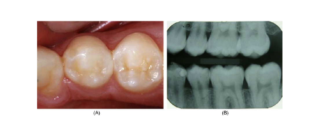
FIGURE 4.26
Figure (A) shows the filling after 1 week and the final X-ray (B) with radiopacity for further radiographic control.
4.10
CONCLUSIONS
In dentistry, nanofillers could be considered an inorganic particle with average size of 40 nm or
0.04 μm. This size, however, is not an innovation in dental composites, because the minifilled com-
posites had the same 0.04 μm (40 nm) filler particles since the 1970s. The real innovation is the nano-
filler manufacture and the possibility of improving the load of inorganic phase
[21]
. The effect of
this high filler load is widely recorded in terms of mechanical properties. Microhybrid composites
with additional load of nanofillers are the best clinical choice. The next research frontier in operative
dentistry is the manufacture of a filler material with shape and composition that can closely mimic the
optical and mechanical characteristics of the natural hard tissues (enamel and dentin).
Acknowledgments
The authors would like to thank the associated professors Prof. Dr Hugo Mistuo Silva Oshima and
Prof. Dr Luciana Mayumi Hirakata and the students of Dentistry Graduate Program of Pontifical
Catholic University of Rio Grande do Sul, Brazil.
References
[1] R.L. Bowen, Properties of a silica-reinforced polymer for dental restorations, J. Am. Dent. Assoc. 66 (1963)
57-64.
[2] Y. Xia, F. Zhang, H. Xie, N. Gu, Nanoparticle-reinforced resin-based dental composites, J. Dent. 36 (6)
(2008) 450-455.
[3] S.B. Mitra, D. Wu, B.N. Holmes, An application of nanotechnology in advanced dental materials, J. Am.
Dent. Assoc. 134 (2003) 1382-1390.