Biomedical Engineering Reference
In-Depth Information
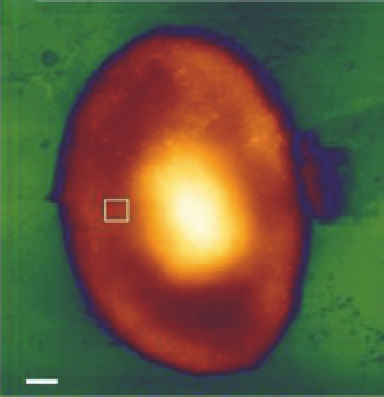
(A)
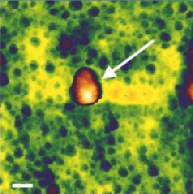
(B)
100 nm
1
µ
m
FIGURE 21.14
Influenza virus particles associated with the surface of an erythrocyte. The overview image (A) and
higher-magnification image (B) of the red blood cell surface show influenza virus particles (white
arrow)
[20]
.
are specifically captured from a sample solution on the surface of our sensor
[21].
This is illustrated
in
Figure 21.15
.
21.8
NANODENTISTRY
There are significant advances in dentistry that illuminated the road for the shift from “macro” to
“nano” in dental sciences. It is evident that increases in the versatility of scientific knowledge and
the ability to control physical processes at a finer resolution naturally lead to more information and,
henceforth, to more questions. The broader our knowledge, the more amazement arises in face of nat-
ural wonders
[22,23]
. The same could certainly be said for the field of dentistry. The historic progress
in this area naturally goes hand in hand with many new questions and challenges that provide oppor-
tunities for improvement. The progress, admittedly, has been slower than might be considered desir-
able for those who would wish to put a cutting-edge technology to clinical use. For example, early
descriptions of the extraction of teeth with the use of forceps by Hippocrates and Aristotle date back
to 500-300 BC, a technique that has remained essentially unchanged till date. Likewise, restorations
with amalgam and gold date back to years 700 and 1746, respectively, and are still a part of our clini-
cal setting without much change.
21.8.1
The Impact of Nanotechnology
It is, on the other hand, valid to point out that nanotechnology slowly had made its way from the
lab bench to any other technological or medical field. This is hampered not only by slow progress