Biomedical Engineering Reference
In-Depth Information
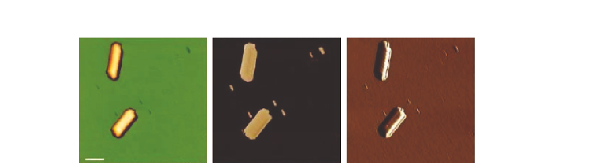
(A)
(B)
(C)
200 nm
FIGURE 21.13
Intermittent contact mode images of TMV particles. Topography (A), phase (B), and error signal
images (C) of the same sample region are presented
[20]
.
to propagate. Viruses are extremely small, around 20-400 nm. This means that the characterization
of viral structure requires high-resolution imaging techniques, such as AFM. Here TMV has been
imaged, the first virus imaged using electron microscopy.
The TMV particles were adsorbed to mica and imaged using intermittent contact mode. These
virus particles are known as helical capsids, where the coat protein stacks in a helical pattern around
the genetic material. This helical stacking can be seen in the height, phase, and error signal chan-
nels (
Figure 21.13
). One significant advantage of using a BioAFM, such as the JPK Nanowizard®, to
image biological samples is that the imaging can be conducted in fluid. As such, virus particles can be
imaged on the surface of their target cells in fluid. Here, influenza virus has been imaged, attached to
the surface of red blood cells, in fluid, using intermittent contact mode. The virus particles are clearly
imaged at the surface of the cells (
Figure 21.14
).
21.7
NANOPLASMONIC SENSORS DETECTING LIVE VIRUSES
The recent emergence of H1N1 and H5N1 flu viruses and severe acute respiratory syndrome has
highlighted the importance of rapid detection and accurate diagnosis in health-care and preventive
medicine. The problem in these areas is that many virus detection platforms have limitations because
they are not easily compatible with point-of-care use without the existence of significant infrastruc-
ture. Cell culturing is a time-consuming, highly specialized, and labor-intensive process. Therefore,
highly sensitive/specific, compact, rapid, and easy to use virus diagnostics are needed to prevent fur-
ther spread at the onset of a viral epidemic.
Unlike techniques based on external labeling, such resonance shifting operates as a reporter of the
molecular binding phenomena in a label-free fashion and enables transduction of the capturing event
directly to the far field optical signal. Specific detection of viruses in a label-free fashion requires an
effective method to distinguish nonspecific binding of the viruses to the plasmonic sensor surface.
Selectivity is achieved by surface immobilized highly specific antiviral immunoglobulins showing
strong affinity to the viral membrane proteins. Correspondingly, with the use of antibodies, viruses