Biomedical Engineering Reference
In-Depth Information
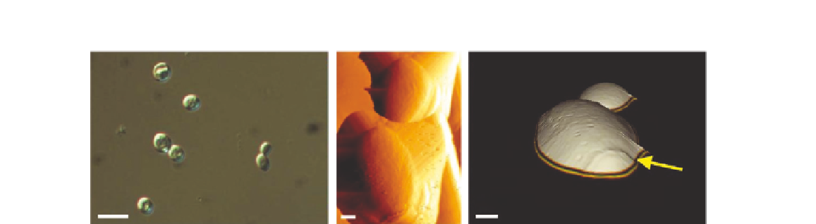
(A)
(B)
(C)
10
µ
m
1
µ
m
1
µ
m
FIGURE 21.9
Imaging of
S. cerevisiae
. Yeast cells were located using DIG microscopy (A) and then imaged in contact mode
in fluid with the AFM (B). In (C) a 3D image generated from the height channel is displayed, highlighting the
bud scar on the mother cell (white arrow)
[20]
.
21.6.2
Bacteria
As AFM is a surface imaging technique, it has been used to characterize surface structures at high
resolution. The most commonly used laboratory bacteria is
E. coli
, a gram-negative bacteria. Gram-
negative bacteria have a plasma membrane, surrounded by a periplasmic space in which there is a
rigid but highly porous cell wall of peptidoglycan. This is then surrounded by an outer membrane,
from which lipopolysaccharides of varying length extend.
Here, two strains of
E. coli
, DH5a and OP50, have been imaged. The images of DH5a show the
classic, rod shape of many gram-negative bacteria. The cells were scanned in air (
Figure 21.10
) and
in buffer (
Figure 21.11
). When imaged in air, the surface of the bacteria appears highly patterned. In
addition, a halo around the bacterium is apparent. These structures likely correspond to pill, which are
found at the surface of
E. coli
. In contrast when imaged in fluid (
Figure 21.11
), the surface of the bac-
terium appears much smoother. In this case this is due to the fact that the surface structures would be
easily displaced by the movement of the tip during scanning, as they are not fixed in place.
The OP50 strain, while also
E. coli
, appears quite different. OP50 was originally isolated as a
strain that could be used to feed
Caenorhabditis elegans
. It is a uracil-requiring strain that is more
fragile and smaller than other
E. coli
strains. When imaged in air (
Figure 21.12
), these bacteria do not
exhibit the same structured surface as seen for the DH5a. In addition, the cells are more fragile and
must be carefully imaged to avoid removing them from the surface. In fluid, the surface is also less
structured than that of DH5a, and regions of the cell surface are displaced in the scan direction, likely
corresponding to the displacement of the sugars and other flexible structures at the surface of the cell.
21.6.3
AFM Study of the Structure-Function Relationship of the Biofilm-Forming
Bacterium
Streptococcus mutans
AFM has garnered much interest in recent years for its ability to probe the structure, function,
and cellular nanomechanics inherent to specific biological cells. In particular, AFM has been used
to probe the important structure-function relationships of the bacterium
Streptococcus mutans
.