Biomedical Engineering Reference
In-Depth Information
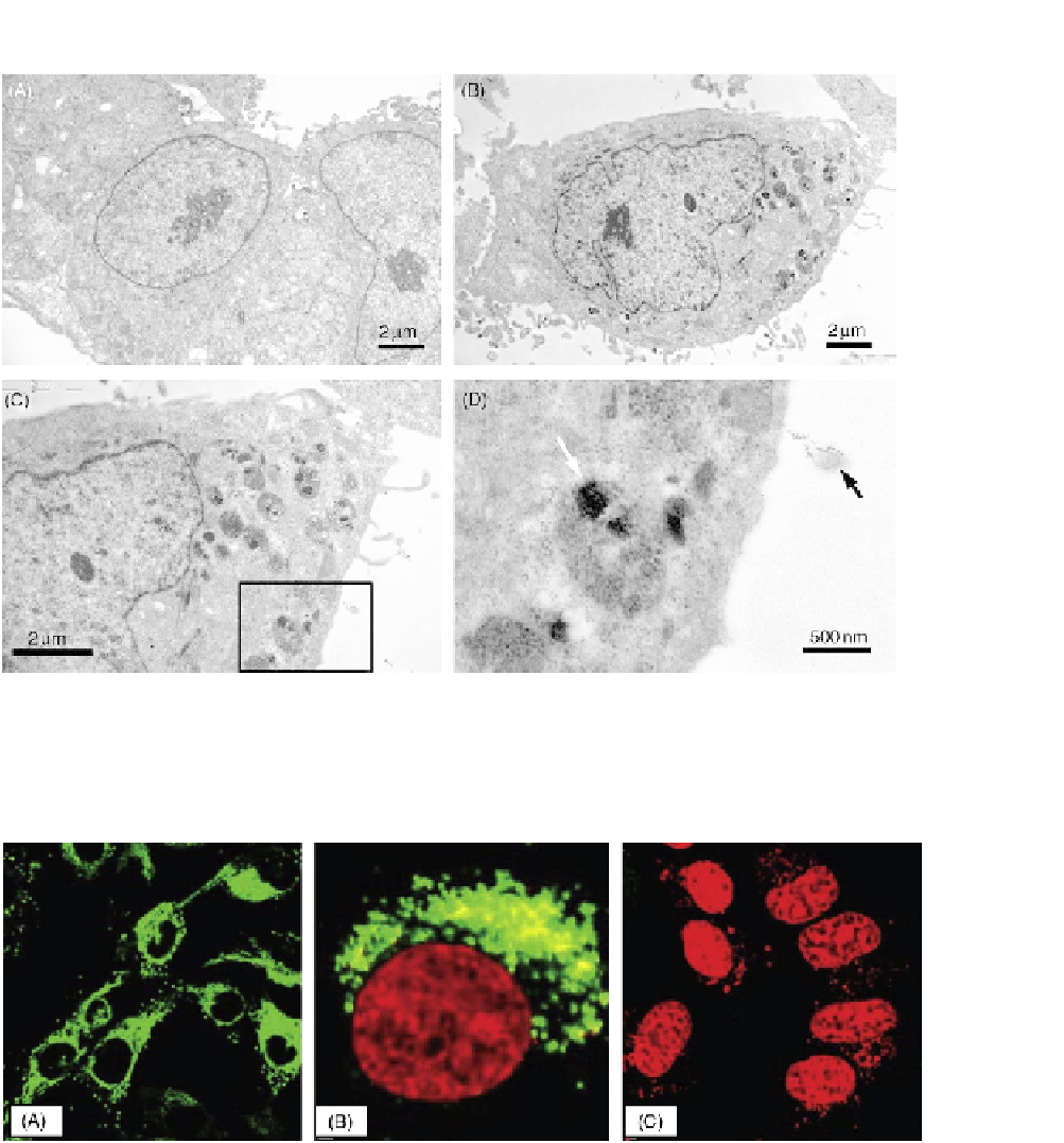
FIGURE 20.7
TEM of (A) HeLa cells before treatment, (B) HeLa cells treated with DOX-FA-CHI-ALG-SWNTs, (C) a
magnified image of (B), (D) magnified image of the boxed region in (C). The black arrow points at a SWNT-
containing vesicle, and the white arrow points at some aggregated nanotubes inside a lysosome
[27]
.
FIGURE 20.8
Confocal microscopy showing the internalization of labeled single-strand DNA into HeLa cell using SWNTs.
(A) The labeled DNA (gray color) is surrounding the nucleus (black circles) at 37°C. (B) The nucleus (large
gray area) is surrounded by the labeled DNA after internalization at 37°C. (C) At 4°C, no DNA internalization
has occurred.