Biomedical Engineering Reference
In-Depth Information
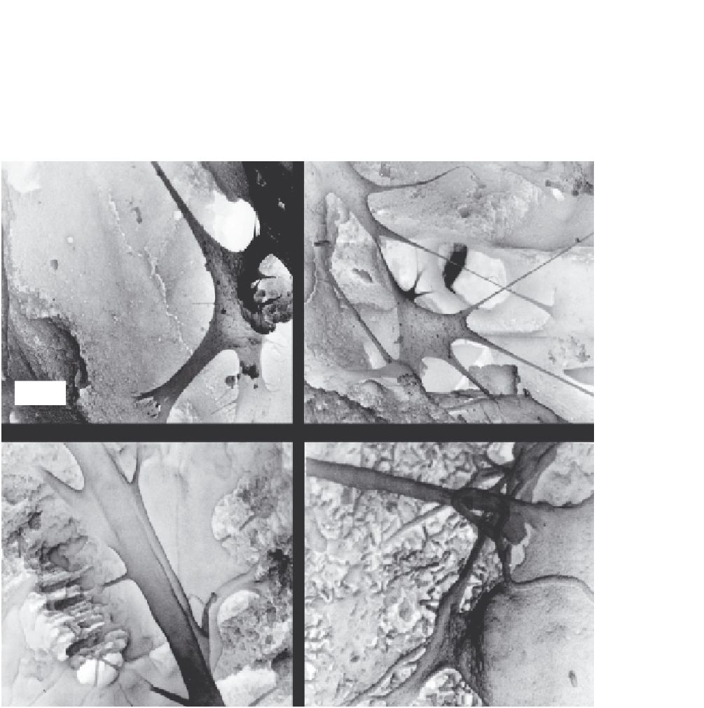
had established cell-cell junction (arrow) in the pore (B). In (C), secondary extensions sprouted from
the primary extension “E.” The cell extensions were shown (D) to have anchored to the nano-apatite
crystals that make up the CPC matrix. Hence, cells were able to infiltrate into the macroporous CPC
and attach to the nano-apatite crystals.
(B)
(A)
50
µ
m
O
O
E
10
µ
m
(C)
(D)
E
E
Nano-apatite
500 nm
1
µ
m
E
FIGURE 12.2
SEM showing cell infiltration into the macropores of CPC. (A) The pore was large enough for the
osteoblastic cell “O” (mouse MC3T3-E1 cells), and the cell had developed cytoplasmic extensions “E.” (B)
Two cells had established cell-cell junction (arrow) in the pore. (C) Secondary extensions sprouting from
the primary extension “E.” (D) Cell extensions anchored to the nano-apatite crystals of CPC, similar to that
of the stem cells in
Figure 12.1
.
Adapted from Ref.
[57]
with permission.